

Elongated oligomers in beta2-microglobulin amyloid assembly revealed by ion mobility spectrometry-mass spectrometry
- PMID: 20351246
- PMCID: PMC2872402
- DOI: 10.1073/pnas.0913046107
Elongated oligomers in beta2-microglobulin amyloid assembly revealed by ion mobility spectrometry-mass spectrometry
Abstract
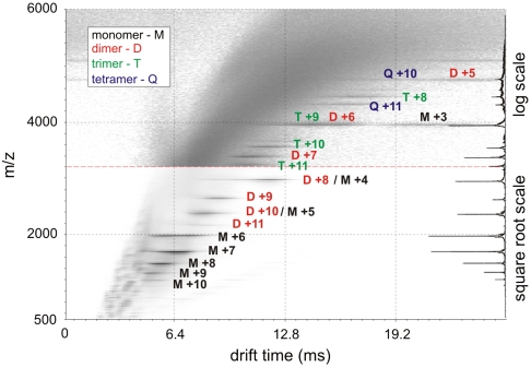
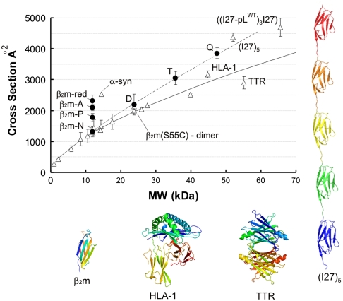
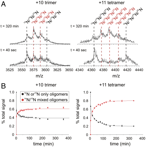
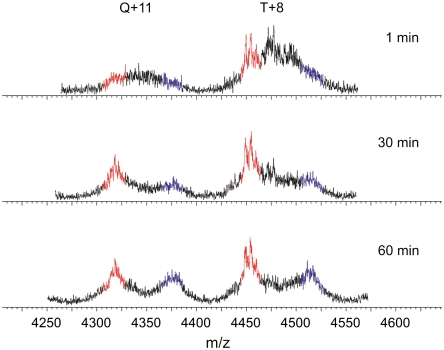
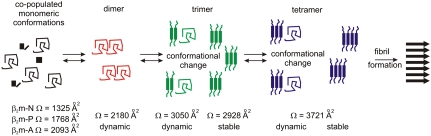
The key to understanding amyloid disease is the characterization of oligomeric species formed during the early stages of fibril assembly. Here we have used electrospray ionisation-ion mobility spectrometry-mass spectrometry to identify and structurally characterize the oligomers formed during amyloid assembly from beta(2)-microglobulin (beta(2)m). Beta(2)m oligomers are shown to have collision cross-sections consistent with monomeric units arranged in elongated assemblies prior to fibril formation. Direct observation, separation, and quantification of transient oligomeric species reveals that monomers to tetramers are populated through the lag phase with no evidence for the significant population of larger oligomeric species under the conditions employed. The dynamics of each oligomeric species were monitored directly within the ensemble at concentrations commensurate with amyloid formation by observing the subunit exchange of (14)N- and (15)N-labeled oligomers. Analysis of the data revealed a decrease in oligomer dynamics concomitant with increasing oligomer size and the copopulation of dynamic dimeric and trimeric species with more stable trimeric and tetrameric species. The results presented map the events occurring during the lag phase of fibril formation and give a clear insight into the structural characteristics and dynamic nature of the beta(2)m oligomers, demonstrating the existence of elongated assemblies arising from an intact amyloidogenic protein during fibril formation.
Conflict of interest statement
The authors declare no conflict of interest.
Figures
Similar articles
-
Structure and dynamics of oligomeric intermediates in β2-microglobulin self-assembly.Biophys J. 2011 Sep 7;101(5):1238-47. doi: 10.1016/j.bpj.2011.07.023. Biophys J. 2011. PMID: 21889462 Free PMC article.
-
Characterization of Amyloid Oligomers by Electrospray Ionization-Ion Mobility Spectrometry-Mass Spectrometry (ESI-IMS-MS).Methods Mol Biol. 2016;1345:115-32. doi: 10.1007/978-1-4939-2978-8_8. Methods Mol Biol. 2016. PMID: 26453209
-
Insights into the role of the beta-2 microglobulin D-strand in amyloid propensity revealed by mass spectrometry.Mol Biosyst. 2014 Mar 4;10(3):412-20. doi: 10.1039/c3mb70420c. Epub 2013 Dec 12. Mol Biosyst. 2014. PMID: 24336936 Free PMC article.
-
Advances in ion mobility spectrometry-mass spectrometry reveal key insights into amyloid assembly.Biochim Biophys Acta. 2013 Jun;1834(6):1257-68. doi: 10.1016/j.bbapap.2012.10.002. Epub 2012 Oct 11. Biochim Biophys Acta. 2013. PMID: 23063533 Free PMC article. Review.
-
Limited proteolysis in the investigation of beta2-microglobulin amyloidogenic and fibrillar states.Biochim Biophys Acta. 2005 Nov 10;1753(1):44-50. doi: 10.1016/j.bbapap.2005.09.004. Epub 2005 Sep 23. Biochim Biophys Acta. 2005. PMID: 16213198 Review.
Cited by
-
Traveling-wave Ion Mobility-Mass Spectrometry Reveals Additional Mechanistic Details in the Stabilization of Protein Complex Ions through Tuned Salt Additives.Int J Ion Mobil Spectrom. 2013 Mar 1;16(1):41-50. doi: 10.1007/s12127-013-0121-9. Epub 2013 Jan 29. Int J Ion Mobil Spectrom. 2013. PMID: 23539363 Free PMC article.
-
Structural Characterization of Monomers and Oligomers of D-Amino Acid-Containing Peptides Using T-Wave Ion Mobility Mass Spectrometry.J Am Soc Mass Spectrom. 2017 Jan;28(1):110-118. doi: 10.1007/s13361-016-1523-9. Epub 2016 Nov 7. J Am Soc Mass Spectrom. 2017. PMID: 27822705 Free PMC article.
-
High-resolution separation of bioisomers using ion cloud profiling.Nat Commun. 2023 Mar 20;14(1):1535. doi: 10.1038/s41467-023-37281-7. Nat Commun. 2023. PMID: 36941278 Free PMC article.
-
Ion Mobility Spectrometry-Mass Spectrometry of Intrinsically Unfolded Proteins: Trying to Put Order into Disorder.Curr Anal Chem. 2013 Apr;9(2):181-191. doi: 10.2174/1573411011309020004. Curr Anal Chem. 2013. PMID: 23885220 Free PMC article.
-
Amyloid-β-neuropeptide interactions assessed by ion mobility-mass spectrometry.Phys Chem Chem Phys. 2013 Jun 21;15(23):8952-61. doi: 10.1039/c3cp50721a. Epub 2013 Apr 24. Phys Chem Chem Phys. 2013. PMID: 23612608 Free PMC article.
References
-
- Gejyo F, et al. A new form of amyloid protein associated with chronic-hemodialysis was identified as beta 2-microglobulin. Biochem Biophys Res Commun. 1985;129:701–706. - PubMed
-
- Kad NM, et al. Hierarchical assembly of beta 2-microglobulin amyloid in vitro revealed by atomic force microscopy. J Mol Biol. 2003;330:785–797. - PubMed
Publication types
MeSH terms
Substances
Grants and funding
LinkOut - more resources
Full Text Sources
Research Materials
