. 2010 Apr 27;107(17):7692-7.
doi: 10.1073/pnas.1002753107. Epub 2010 Mar 29.

ErbB3/HER3 intracellular domain is competent to bind ATP and catalyze autophosphorylation

Affiliations

ErbB3/HER3 intracellular domain is competent to bind ATP and catalyze autophosphorylation

Fumin Shi et al. Proc Natl Acad Sci U S A. .

Abstract

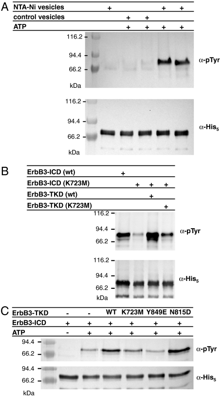
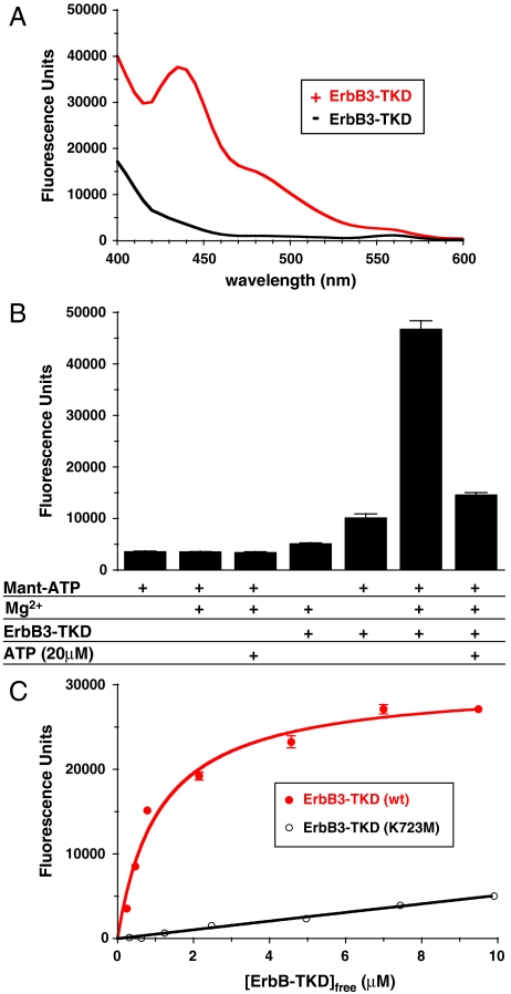
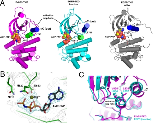
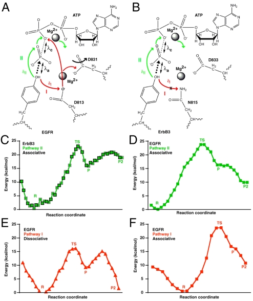
ErbB3/HER3 is one of four members of the human epidermal growth factor receptor (EGFR/HER) or ErbB receptor tyrosine kinase family. ErbB3 binds neuregulins via its extracellular region and signals primarily by heterodimerizing with ErbB2/HER2/Neu. A recently appreciated role for ErbB3 in resistance of tumor cells to EGFR/ErbB2-targeted therapeutics has made it a focus of attention. However, efforts to inactivate ErbB3 therapeutically in parallel with other ErbB receptors are challenging because its intracellular kinase domain is thought to be an inactive pseudokinase that lacks several key conserved (and catalytically important) residues-including the catalytic base aspartate. We report here that, despite these sequence alterations, ErbB3 retains sufficient kinase activity to robustly trans-autophosphorylate its intracellular region--although it is substantially less active than EGFR and does not phosphorylate exogenous peptides. The ErbB3 kinase domain binds ATP with a K(d) of approximately 1.1 microM. We describe a crystal structure of ErbB3 kinase bound to an ATP analogue, which resembles the inactive EGFR and ErbB4 kinase domains (but with a shortened alphaC-helix). Whereas mutations that destabilize this configuration activate EGFR and ErbB4 (and promote EGFR-dependent lung cancers), a similar mutation conversely inactivates ErbB3. Using quantum mechanics/molecular mechanics simulations, we delineate a reaction pathway for ErbB3-catalyzed phosphoryl transfer that does not require the conserved catalytic base and can be catalyzed by the "inactive-like" configuration observed crystallographically. These findings suggest that ErbB3 kinase activity within receptor dimers may be crucial for signaling and could represent an important therapeutic target.


Conflict of interest statement

The authors declare no conflict of interest.

Figures

Fig. 1.
Fig. 1.
Autophosphorylation of the ErbB3 intracellular domain in vitro. (A) ErbB3-ICD (3 μM) becomes autophosphorylated when incubated with ATP and vesicles containing 10% (mol/mol) NTA-Ni lipid (right two lanes), but not when either ATP or NTA-Ni lipid is absent. The upper panel is antiphosphotyrosine blot, and the lower is antipentahistidine loading control. Lanes 3/4 and 5/6 represent duplicate experiments. (B) ErbB3-ICD harboring a K723M mutation shows little autophosphorylation (lane 2) when treated with ATP and NTA-Ni vesicles as in A, whereas wild-type ErbB3-ICD autophosphorylation is robust. His-tagged ErbB3-TKD665–1001 added at 3 μM (lane 3) causes robust trans-phosphorylation of ErbB3-ICD (K723M), which is diminished by a K723M mutation in the TKD (lane 4). (C) Trans-phosphorylation of ErbB3-ICD by wild-type and mutated forms of ErbB3-TKD665–1001 was compared as described in the text.
Fig. 2.
Fig. 2.
Mant-ATP binding to ErbB3-TKD648–1001. (A) Fluorescence emission spectra (with excitation at 280 nm) for 1 μM mant-ATP (plus 5 mM MgCl2) in the absence of ErbB3-TKD (Black) and with 3 μM added ErbB3-TKD648–1001 (Red). (B) Fluorescence emission of 1 μM mant-ATP at 450 nm under different conditions (with excitation at 280 nm). Protein-to-mant FRET is only seen when 1 μM mant-ATP, 5 mM MgCl2, and 3 μM ErbB3-TKD648–1001 are all present. The FRET signal is greatly reduced by adding 20 μM ATP to saturate the nucleotide-binding site in the kinase (far right). (C) Titrating ErbB3-TKD648–1001 into a 0.6 μM mant-ATP solution containing 5 mM MgCl2 yields a hyperbolic binding curve fit to give a Kd value of 1.1 μM for wild-type ErbB3-TKD. Mean ± standard deviation are shown for at least 3 independent experiments in B and C.
Fig. 3.
Fig. 3.
Crystal structure of ErbB3-TKD665–1001. (A) Cartoon representations are shown for ErbB3-TKD665–1001 (Left: Magenta), inactive EGFR-TKD (Middle: Cyan), and active EGFR-TKD (Right: Gray). EGFR-TKD structures are from PDB codes 2GS7 (5) and 2ITX (48). All structures have bound AMP-PNP and Mg2+ (shown in spheres). Helix αC is colored blue in each structure and is denoted as out (ErbB3 and inactive EGFR) or in (active EGFR). The short activation loop helix in ErbB3 and inactive EGFR is colored green. H740 in helix αC of ErbB3 and the similarly located E738 in EGFR are shown as sticks, as are ErbB3 N815 and EGFR D813. β-strands in the N lobe are labeled. (B) Close-up view of Mg2+-AMP-PNP in the ErbB3-TKD665–1001 active site, with electron density shown as a 2Fo - Fc map calculated at 1.8σ by using phases from a model with no nucleotide. Coordination of bound Mg2+ by the N820 and D833 side chains (from the DFG motif) and the AMP-PNP α- and β-phosphates is depicted. N815 (close to the γ-phosphate) is also marked. (C) Close-up of the short activation loop helix seen in ErbB3-TKD665–1001 (Magenta) and inactive EGFR-TKD (Cyan), with the two kinase domains superimposed. Mutations at L834 or L837 in EGFR activate the receptor in NSCLC. These residues overlay with V836 and L839 in ErbB3-TKD665–1001 and interact with the hydrophobic pocket that contributes to stabilization of helix αC in the out position. A V836A mutation inhibits ErbB3-TKD activity.
Fig. 4.
Fig. 4.
QM/MM simulations of phosphoryl transfer. Schematic of the phosphoryl-transfer pathway in (A) EGFR-TKD and (B) ErbB3-TKD, as captured in QM/MM simulations. Mg2+ ions are marked, as are the catalytic aspartates (D831 in EGFR, D833 in ErbB3), the proposed catalytic base in EGFR (D813), and its replacement in ErbB3 (N815). Two potential pathways for proton migration (concomitant with phosphoryl transfer) are marked as described in the text: pathway I (Red) and pathway II (Green). The distance from the substrate -OH proton to the Oδ2 oxygen of D813 in EGFR is labeled δI, and its distance to the O1γ oxygen of ATP is δII (see Fig. S5 A and B). The nucleophilic attack distance λa (distance between the tyrosine oxygen and the ATP Pγ) and the bond cleavage distance λc (distance between the ATP Pγ and ATP O2/3β) are marked (see Fig. S5). (CF) Energy changes along the reaction pathway in QM/MM simulations for the noted mechanisms involving pathway II (Green) or pathway I (Red). States correspond to “R” (reactant); “TS” (transition state with trigonal-bipyramidal geometry around Pγ); “P” (product after phosphoryl transfer with proton bound to ATP O1γ); and “P2” (product with proton transferred to ATP O2β); see Figs. S4S6 for details. (C) Energy changes along the simulated ErbB3 reaction pathway (in the structure shown in Fig. 3A). Symbols bounded by black squares represent the forward scan, and those bounded by gray squares represent the reverse scan. Only pathway II is utilized, and estimated Ea for phosphoryl transfer is 23 kcal/mol. (D) Energy changes along the EGFR reaction pathway (active configuration) utilizing pathway II and the associative mechanism: Estimated Ea is 24 kcal/mol. (E) Phosphoryl transfer catalyzed by EGFR via the dissociative mechanism (utilizing pathway I for proton migration—via D813) has the lowest Ea value, at 16 kcal/mol. (F) Associative phosphoryl transfer concomitant with pathway I for EGFR has an estimated Ea of 24 kcal/mol.

Comment in

  • Yet another "active" pseudokinase, Erb3.
    Taylor SS, Kornev AP. Taylor SS, et al. Proc Natl Acad Sci U S A. 2010 May 4;107(18):8047-8. doi: 10.1073/pnas.1003436107. Epub 2010 Apr 26. Proc Natl Acad Sci U S A. 2010. PMID: 20421461 Free PMC article. No abstract available.

References

    1. Hynes NE, MacDonald G. ErbB receptors and signaling pathways in cancer. Curr Opin Cell Biol. 2009;21:177–184. - PubMed
    1. Baselga J, Swain SM. Novel anticancer targets: Revisiting ERBB2 and discovering ERBB3. Nat Rev Cancer. 2009;9:463–475. - PubMed
    1. Jura N, et al. Mechanism for activation of the EGF receptor catalytic domain by the juxtamembrane segment. Cell. 2009;137:1293–1307. - PMC - PubMed
    1. Red Brewer M, et al. The juxtamembrane region of the EGF receptor functions as an activation domain. Mol Cell. 2009;34:641–651. - PMC - PubMed
    1. Zhang X, Gureasko J, Shen K, Cole PA, Kuriyan J. An allosteric mechanism for activation of the kinase domain of epidermal growth factor receptor. Cell. 2006;125:1137–1149. - PubMed

Publication types

Associated data