. 2010 Apr 13;107(15):6852-7.
doi: 10.1073/pnas.1000232107. Epub 2010 Mar 29.

Structure of lactococcal phage p2 baseplate and its mechanism of activation

Affiliations

Structure of lactococcal phage p2 baseplate and its mechanism of activation

Giuliano Sciara et al. Proc Natl Acad Sci U S A. .

Abstract

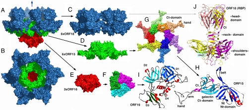
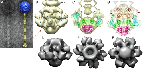
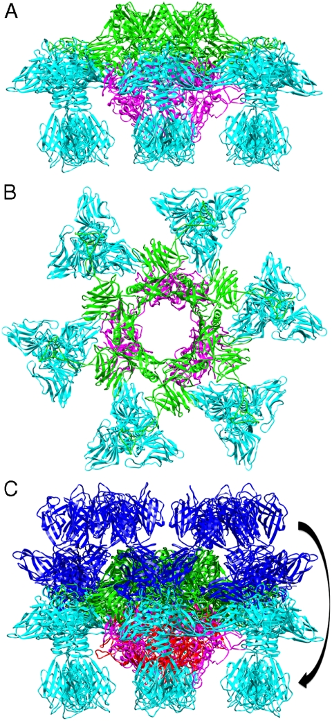
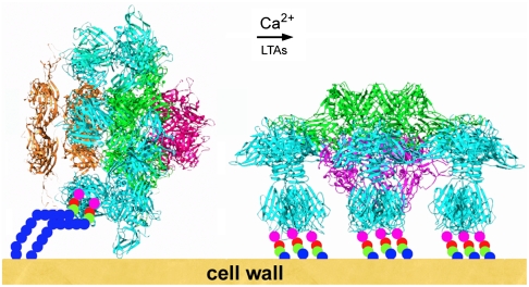
Siphoviridae is the most abundant viral family on earth which infects bacteria as well as archaea. All known siphophages infecting gram+ Lactococcus lactis possess a baseplate at the tip of their tail involved in host recognition and attachment. Here, we report analysis of the p2 phage baseplate structure by X-ray crystallography and electron microscopy and propose a mechanism for the baseplate activation during attachment to the host cell. This approximately 1 MDa, Escherichia coli-expressed baseplate is composed of three protein species, including six trimers of the receptor-binding protein (RBP). RBPs host-recognition domains point upwards, towards the capsid, in agreement with the electron-microscopy map of the free virion. In the presence of Ca(2+), a cation mandatory for infection, the RBPs rotated 200 degrees downwards, presenting their binding sites to the host, and a channel opens at the bottom of the baseplate for DNA passage. These conformational changes reveal a novel siphophage activation and host-recognition mechanism leading ultimately to DNA ejection.


Conflict of interest statement

The authors declare no conflict of interest.

Figures

Fig. 1.
Fig. 1.
Crystal structure of the phage p2 baseplate and its components. (A) View of the baseplate surface; ORF15 is in green, ORF16 in red, and ORF18 in blue. The blue arrow indicates the position of the quasi 6-fold axis and points toward the rest of the phage tail and the capsid. (B) The baseplate has been rotated by 90° around the horizontal axis. The central channel formed by ORF15 hexamer is closed by the ORF16 trimeric dome. (C) The arrangement of ORF18 as six trimers. (D) Hexameric ORF15. (E) Trimeric ORF16. (F) View of ORF16 trimer rotated 180° relative to baseplate (B) with a different color for each subunit. (G) ORF15 hexamer is viewed in the same orientation as in (B). Each subsubunit, as well as the N- and C-terminus domains, have a different color. The central channel is ∼40  wide. (H) Ribbon view of ORF15 subunit. The N- and C-terminus domains have their β-strands colored dark and light blue, respectively; helices are colored red and violet, respectively; and coiled sections are gray. The striking features of the structure, the arm (which helps hexamerization) and the extension (which grips ORF18 trimer) have been identified. Note the closure of the surface at the center of the trimer. (I) Ribbon view of ORF16 subunit. The four domains have been identified by D 1 to 4 and different colors of the β-strands. Domain 4 is only helical. These domains correspond to those identified in gp27 from phage T4 (12). (J) Ribbon view of the receptor-binding protein ORF18 trimer, with a different color for each chain. The trimer domains of shoulders, neck, and head are represented. The VHHs are displayed in Fig. S2.
Fig. 2.
Fig. 2.
EM structure of the baseplate of phage p2 and results of the fitting of the baseplate crystal structure. (A) EM micrograph of the virulent lactococcal phage p2. Overall EM structure of phage p2 (right) showing the capsid (blue, top), the tail, formed of rings of the major tail protein hexamers (gold), and the globular baseplate (gold, bottom). ( B) View of the EM structure of the baseplate with the first major tail protein (MTP) hexamer. The RBP positions are identified by blue arrows or a blue dot. The RBP head is pointing upwards. (C) View of the crystal structure of the baseplate fitted in the EM map. ORF15 is green, ORF16 red, and ORF18 blue. Each RBP, ORF15 hexamer, and ORF16 trimer have been fitted as rigid blocks. (D), (E), (F) Views of the cryoEM map of the expressed p2 baseplate, from side (D), up (E), and down (F), respectively. (G) Sliced view showing the fitting of a second ORF15 hexamer (brown) in the EM map, 180° rotated relative to that in the crystal structure. The ring formed by the N-terminus has been fitted as a block, and the rotated and translated galectin domains have been fitted as a second block. Note the extension touching the RBP head domain (red arrows).
Fig. 3.
Fig. 3.
The crystal structure of the “heads-down” conformations of p2 baseplate. (A) Side view in ribbon representation of the “heads-down” conformations of p2 baseplate. (B) View from top (ORF15, green; ORF16, pink; ORF18, blue). (C) Superposition of the rings formed by the N-terminal domains of ORF15. ORF18 trimers have undergone a 200° rotation downwards.
Fig. 4.
Fig. 4.
Schematic representation of the putative adsorption mechanism of phage p2 to its host. The rest form (left) of the baseplate is activated by Ca2+ cations and by the traction of lipoteichoic acids bound to a few receptor-binding sites which destabilize the RBPs. They subsequently rotate by 200° and become available for a complete binding. Concomitantly, the tip of the baseplate opens, giving way to dsDNA.

References

    1. Rousseau GM, Moineau S. Evolution of Lactococcus lactis phages within a cheese factory. Appl Environ Microbiol. 2009;75:5336–5344. - PMC - PubMed
    1. Deveau H, Labrie SJ, Chopin MC, Moineau S. Biodiversity and classification of lactococcal phages. Appl Environ Microbiol. 2006;72:4338–4346. - PMC - PubMed
    1. Maniloff J, Ackermann HW. Taxonomy of bacterial viruses: Establishment of tailed virus genera and the order Caudovirales. Arch Virol. 1998;143:2051–2063. - PubMed
    1. Boulanger P, et al. Phage T5 straight tail fiber is a multifunctional protein acting as a tape measure and carrying fusogenic and muralytic activities. J Biol Chem. 2008;283:13556–13564. - PubMed
    1. Plisson C, et al. Structure of bacteriophage SPP1 tail reveals trigger for DNA ejection. Embo J. 2007;26:3720–3728. - PMC - PubMed

Publication types