. 2010 Jun;151(6):2846-57.
doi: 10.1210/en.2009-1032. Epub 2010 Mar 29.

Luteinizing hormone stimulates mammalian target of rapamycin signaling in bovine luteal cells via pathways independent of AKT and mitogen-activated protein kinase: modulation of glycogen synthase kinase 3 and AMP-activated protein kinase

Affiliations

Luteinizing hormone stimulates mammalian target of rapamycin signaling in bovine luteal cells via pathways independent of AKT and mitogen-activated protein kinase: modulation of glycogen synthase kinase 3 and AMP-activated protein kinase

Xiaoying Hou et al. Endocrinology. 2010 Jun.

Abstract

LH stimulates the production of cAMP in luteal cells, which leads to the production of progesterone, a hormone critical for the maintenance of pregnancy. The mammalian target of rapamycin (MTOR) signaling cascade has recently been examined in ovarian follicles where it regulates granulosa cell proliferation and differentiation. This study examined the actions of LH on the regulation and possible role of the MTOR signaling pathway in primary cultures of bovine corpus luteum cells. Herein, we demonstrate that activation of the LH receptor stimulates the phosphorylation of the MTOR substrates ribosomal protein S6 kinase 1 (S6K1) and eukaryotic translation initiation factor 4E binding protein 1. The actions of LH were mimicked by forskolin and 8-bromo-cAMP. LH did not increase AKT or MAPK1/3 phosphorylation. Studies with pathway-specific inhibitors demonstrated that the MAPK kinase 1 (MAP2K1)/MAPK or phosphatidylinositol 3-kinase/AKT signaling pathways were not required for LH-stimulated MTOR/S6K1 activity. However, LH decreased the activity of glycogen synthase kinase 3Beta (GSK3B) and AMP-activated protein kinase (AMPK). The actions of LH on MTOR/S6K1 were mimicked by agents that modulated GSK3B and AMPK activity. The ability of LH to stimulate progesterone secretion was not prevented by rapamycin, a MTOR inhibitor. In contrast, activation of AMPK inhibited LH-stimulated MTOR/S6K1 signaling and progesterone secretion. In summary, the LH receptor stimulates a unique series of intracellular signals to activate MTOR/S6K1 signaling. Furthermore, LH-directed changes in AMPK and GSK3B phosphorylation appear to exert a greater impact on progesterone synthesis in the corpus luteum than rapamycin-sensitive MTOR-mediated events.


Figures

Figure 1
Figure 1
LH stimulates time- and concentration-dependent phosphorylation of S6K1 at Thr389 and stimulates S6K1 activity. A, Bovine luteal cells were treated for 15 min with increasing concentrations (1–100 ng/ml) of LH. Western blot analysis was performed using antibodies against the phosphospecific S6K1(Thr389) (1:1000) and total S6K1 (1:000). B, Quantitative analysis of concentration-response relationships as described in A. Data are shown as a percentage of the maximal response to LH in each experiment. Shown are means ± sem from three separate experiments. *, P < 0.05, first statistically significant increase. C, Cells were treated with LH (100 ng/ml) for 5, 15, 30, 60, and 120 min. After the treatment, the cells were lysed and subjected to Western blot analysis using antibodies against the phosphospecific S6K1(Thr389). ACTB protein (anti-ACTB) was used as a loading control. D, Luteal cells were treated with LH (100 ng/ml), 8-Br-cAMP (1 mm), or forskolin (10 μm) for 15 min. Western blot analysis was performed using phosphospecific antibodies against S6K1(Thr389), S6K1(Thr421/Ser424) and RPS6(Ser234/235) (1:1000). ACTB protein (anti-ACTB) was used as a loading control. E, Luteal cells were treated with control medium (CTL), LH (100 ng/ml), 8-Br-cAMP (1 mm), or FBS (10%) for 15 min. S6K1 antibodies or nonimmune serum [normal rabbit serum (NRS)] was used for immunoprecipitation, and S6K1 activity was determined using a GST-S6 fusion protein as substrate. GST alone served as a negative control. Shown are an autoradiogram of radioactive GST-S6 (top row) and a Western blot of the immunoprecipitated S6K1 (bottom row). F, Cells were treated as described in D, and S6K1 protein kinase activity was expressed relative to the activity observed in control samples in each experiment. Data are means ± sem from three separate experiments. *, P < 0.05 vs. CTL.
Figure 2
Figure 2
LH-stimulated S6K1 activation does not involve PI3K or MAK2K/MAPK1/3. A, Bovine luteal cells were pretreated with vehicle, U0126 (20 μm), or wortmannin (100 nm) for 60 min before treatment with control medium (CTL), LH (100 ng/ml), or IGF-I (50 ng/ml) for 15 min. Western blot analysis was performed using phosphospecific antibodies S6K1(Thr389) (1:1000), AKT(Ser 473) (1:1000), MAPK1/3(Thr202/Tyr204) (1:5000), and RPS6(Ser234/235) (1:1000). AKT, MAPK1/3, and ACTB proteins were used as controls. B, Quantitative analysis of phospho-S6K1(Thr389) in the experimental approach described in A. Data are shown as fold increases over the untreated control samples in each experiment. Shown are means ± sem from three separate experiments. Bars with different letters are statistically different at P < 0.05. C, Luteal cells were treated with control medium (CTL), LH (100 ng/ml), or IGF-I (50 ng/ml) for 15 min. Western blot analysis was performed to determine the phosphorylation of TSC2(Thr1462) (1:1000), AKT(Ser473), PRAS40(Thr246) (1:1000), and MTOR(Ser2448) (1:000). D, Summary of experiments described in C. Data are shown as fold increases over the untreated control samples in each experiment. Shown are means ± sem from three separate experiments. *, P < 0.05 vs. control; **, P < 0.05 IGF, vs. LH.
Figure 3
Figure 3
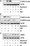
Rapamycin inhibits LH- and FSK-stimulated S6K1 phosphorylation as well as LH- and 8-Br-cAMP-induced cap-dependent translation. A, Bovine luteal cells were pretreated with or without rapamycin (20 nm) for 60 min before treatment for 15 min with control medium, LH (100 ng/ml), or forskolin (10 μm). Western blot analysis was used to determine phosphorylation of S6K1 and RPS6 using antibodies described in Fig. 1. Results are representative of three experiments. B, Luteal cells were pretreated with control medium or rapamycin (20 nm) for 1 h before treatment for 15 min with medium (CTL), FBS (10%), or LH (100 ng/ml). Western blot analysis was performed to determine phosphorylation of S6K1 and 4EBP1(Ser65) (1:1000). C, Luteal cells were pretreated with control medium or rapamycin (20 nm) for 1 h before treatment for 15 min with medium (CTL), 8-Br-cAMP (1 mm), or LH (100 ng/ml). The eIF4E-containing complexes were precipitated using m7G-Sepharose and separated on 13% SDS-PAGE gels. Western blot analysis was performed using antibody to detect coprecipitating 4EBP1 (1:1000) and eIF4E (1:000).
Figure 4
Figure 4
Deactivating GSK3B with LiCl or GSKi, GSK3B inhibitors, induced S6K1 and RPS6 phosphorylation. A, Bovine luteal cells were treated with medium (CTL) or LH (100 ng/ml) for 15 min. Western blot analysis was performed with phosphospecific GSK3B(Ser9) antibodies (1:2000) or total GSK3B antibodies (1:000). ACTB protein was used as a loading control. B, Luteal cells were treated with medium (CTL) or LH (100 ng/ml) for up to 60 min. Western blot analysis was performed and results expressed as a ratio of phospho-GSK3B(Ser9) to ACTB in each experiment. Results are expressed as mean ± sem of three separate experiments. C, Luteal cells were treated with GSKi (10 μm) or LiCl (20 mm) for 15, 60, and 90 min. Western blot analysis was performed to determine the phosphorylation of S6K1(Thr389) as described in Fig. 1.
Figure 5
Figure 5
LH treatment inhibits AMPK activity and ACACA phosphorylation, whereas AICAR inhibits LH-stimulated S6K1 phosphorylation. Bovine luteal cells were treated without (CTL) or with LH (100 ng/ml) for 15 min. The cells were lysed and Western blot analysis was performed using phosphospecific antibodies AMPKα subunit (Thr172) (1:1000), AMPKα subunit (Ser485) (1:1000), ACACA(Ser79) (1:1000), and GSK3B(Ser9). Total AMPKα subunit (1:000) and ACTB were also determined. Densitometry readings were obtained and expressed as a ratio of LH to CTL. B and C, Bovine luteal cells were treated without (CTL) or with LH (100 ng/ml) for 15 min. Western blot analysis was performed to determine levels of AMPKα(Thr172)and ACACA(Ser79). Total AMPKα subunit (1:000) and ACTB were also determined. Data are presented as a percentage of the levels observed in the control samples in each experiment. Results are expressed as mean ± sem in three experiments. *, P < 0.05, LH vs. control. D, Luteal cells were pretreated with AICAR (1 mm) for 60 min before treatment for 15 min with control medium or LH (100 ng/ml). Western blot analysis was conducted to determine ACACA and S6K1 phosphorylation using antibodies described above. ACTB was used as a loading control.
Figure 6
Figure 6
Rapamycin does not block LH-induced progesterone synthesis. Bovine luteal cells were treated with or without LH (100 ng/ml) in the presence or absence with rapamycin (20 nm) (A) or AICAR (1 mm) (B) for 6 h. The conditioned medium was collected, and progesterone levels were determined by RIA. Results are presented as mean ± sem of three or more experiments each conducted in triplicate. Bars with different letters are statistically different at P < 0.05.
Figure 7
Figure 7
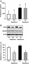
Rapamycin treatment for 24 h does not block LH-induced progesterone synthesis. Bovine luteal cells were treated with or without rapamycin (20 nm) for 24 h. The cells were washed, medium with or without rapamycin was replaced, and then the cells were treated with control medium (CTL) or LH (100 ng/ml) for an additional 6 h. A, The conditioned media were collected for progesterone analysis. B, The cells were also collected and lysed for Western blot analysis of RPS6, HSD3B, and ACTB. C, Levels of RPS6 are presented relative to the level observed in control samples in each experiment. Results are expressed as mean ± sem; n = 4. Bars with different letters are statistically different at P < 0.05.
Figure 8
Figure 8
Proposed model of LH-mediated regulation of MTOR signaling in luteal cells. The results described are consistent with the model in which LH activates the LH receptor and adenylyl cyclase, which leads to an increase in cAMP. As a result, there is an increase in the phosphorylation and inactivation of GSK3B. At the same time, there is a reduction in the activation of AMPK. These events reduce the activity of the TSC, allowing MTOR to phosphorylate components of the translational apparatus, S6K1 and 4EBP1. Treatment with rapamycin inhibits MTOR activation without altering progesterone secretion. Treatment with AICAR activates AMPK, inhibits MTOR phosphorylation of S6K1, and inhibits progesterone secretion. The results suggest that LH uses a unique pathway to activate MTOR signaling in bovine luteal cells, but the rapamycin-sensitive MTOR signaling pathway does not contribute to corpus luteum progesterone synthesis.

Similar articles

Cited by

References

    1. Berisha B, Schams D 2005 Ovarian function in ruminants. Domest Anim Endocrinol 29:305–317 - PubMed
    1. Davis JS, Rueda BR 2002 The corpus luteum: an ovarian structure with maternal instincts and suicidal tendencies. Front Biosci 7:d1949–d1978 - PubMed
    1. Stocco C, Telleria C, Gibori G 2007 The molecular control of corpus luteum formation, function, and regression. Endocr Rev 28:117–149 - PubMed
    1. Marsh JM 1976 The role of cyclic AMP in gonadal steroidogenesis. Biol Reprod 14:30–53 - PubMed
    1. Niswender GD 2002 Molecular control of luteal secretion of progesterone. Reproduction 123:333–339 - PubMed

Publication types

MeSH terms