

Systemic spironucleosis in 2 immunodeficient rhesus macaques (Macaca mulatta)
- PMID: 20351359
- PMCID: PMC3046777
- DOI: 10.1177/0300985810363704
Systemic spironucleosis in 2 immunodeficient rhesus macaques (Macaca mulatta)
Abstract
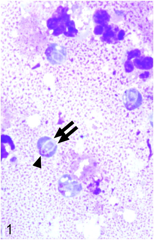
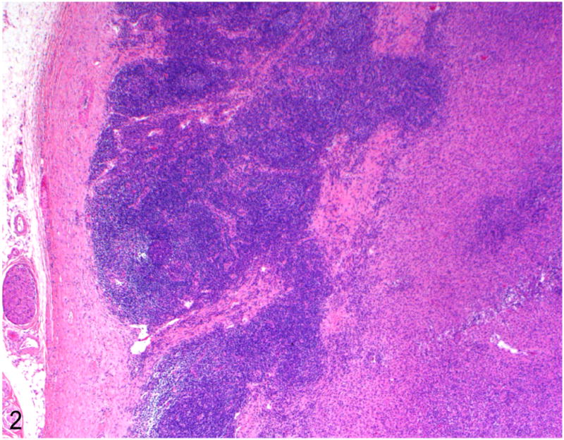
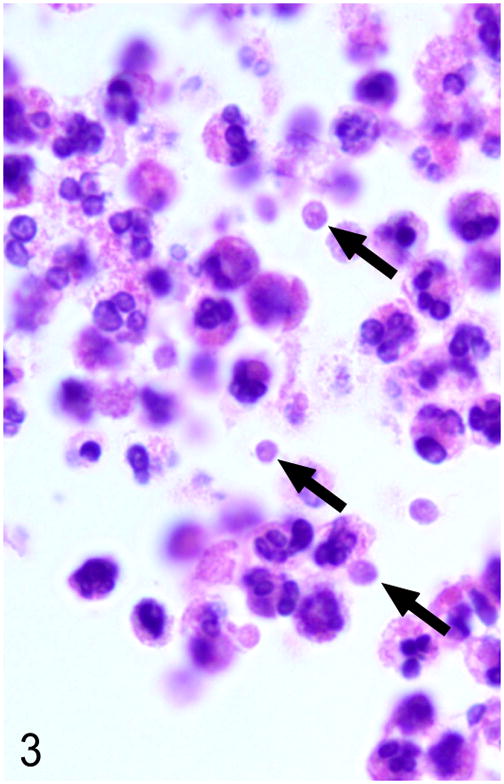
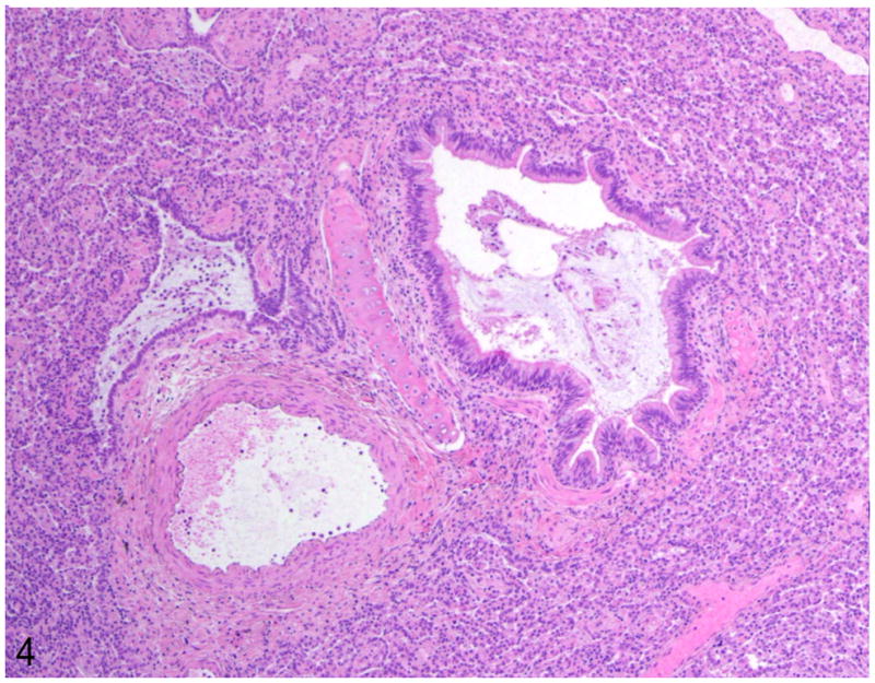
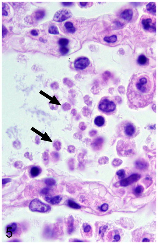
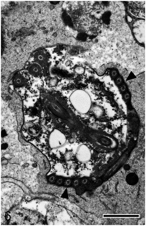
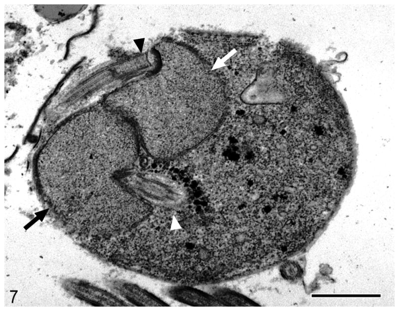
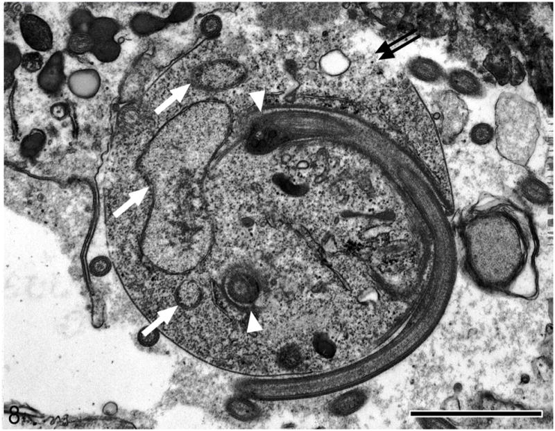
Spironucleus spp are parasites of fish and terrestrial vertebrates, including mice and turkeys, that rarely cause extraintestinal disease. Two rhesus macaques (Macaca mulatta) were experimentally inoculated with simian immunodeficiency virus mac251. Both progressed to simian acquired immune deficiency syndrome within 1 year of inoculation and developed systemic protozoal infections in addition to common opportunistic infections, including rhesus cytomegalovirus, rhesus lymphocryptovirus, and rhesus adenovirus. In the first case, the protozoa were associated with colitis, multifocal abdominal abscessation, and lymphadenitis. In the second case, they were one of a number of organisms associated with extensive pyogranulomatous pneumonia and colitis. Ultrastructural, molecular, and phylogenetic analysis revealed the causative organism to be a species of Spironucleus closely related to Spironucleus meleagridis of turkeys. This report is the first of extraintestinal infection with Spironucleus sp in higher mammals and expands the list of opportunistic infections found in immunocompromised rhesus macaques.
Figures

Similar articles
-
Mesenchymoproliferative enteropathy associated with dual simian polyomavirus and rhesus cytomegalovirus infection in a simian immunodeficiency virus-infected rhesus macaque (Macaca mulatta).Vet Pathol. 2013 Jul;50(4):715-21. doi: 10.1177/0300985812463405. Epub 2012 Oct 9. Vet Pathol. 2013. PMID: 23051916 Free PMC article.
-
Ultrastructural morphology of Enterocytozoon bieneusi in biliary epithelium of rhesus macaques (Macaca mulatta).Vet Pathol. 1998 Jul;35(4):292-6. doi: 10.1177/030098589803500408. Vet Pathol. 1998. PMID: 9684973
-
Localization of persistent Enterocytozoon bieneusi infection in normal rhesus macaques (Macaca mulatta) to the hepatobiliary tree.J Clin Microbiol. 1998 Aug;36(8):2336-8. doi: 10.1128/JCM.36.8.2336-2338.1998. J Clin Microbiol. 1998. PMID: 9666017 Free PMC article.
-
[The immunological manifestations and cytological characteristics of infection caused by the simian immunodeficiency virus (SIV) in rhesus monkeys].Tsitol Genet. 1993 Nov-Dec;27(6):97-104. Tsitol Genet. 1993. PMID: 8066812 Review. Russian.
-
Comparative pathobiology of HIV- and SIV-associated lymphoma.AIDS Res Hum Retroviruses. 2001 May 20;17(8):745-51. doi: 10.1089/088922201750237013. AIDS Res Hum Retroviruses. 2001. PMID: 11429114 Review. No abstract available.
Cited by
-
Single-dose bNAb cocktail or abbreviated ART post-exposure regimens achieve tight SHIV control without adaptive immunity.Nat Commun. 2020 Jan 7;11(1):70. doi: 10.1038/s41467-019-13972-y. Nat Commun. 2020. PMID: 31911610 Free PMC article.
-
Spironucleus muris and Eperythrozoon coccoides in Rodents from Northwestern Iran: Rare Infections.J Arthropod Borne Dis. 2018 Dec 25;12(4):334-340. eCollection 2018 Dec. J Arthropod Borne Dis. 2018. PMID: 30915373 Free PMC article.
-
Research Relevant Conditions and Pathology in Nonhuman Primates.ILAR J. 2020 Dec 31;61(2-3):139-166. doi: 10.1093/ilar/ilab017. ILAR J. 2020. PMID: 34129672 Free PMC article. Review.
-
Oxygen induces the expression of invasion and stress response genes in the anaerobic salmon parasite Spironucleus salmonicida.BMC Biol. 2019 Mar 1;17(1):19. doi: 10.1186/s12915-019-0634-8. BMC Biol. 2019. PMID: 30823887 Free PMC article.
-
Immune perturbation following SHIV infection is greater in newborn macaques than in infants.JCI Insight. 2024 Aug 27;9(19):e144448. doi: 10.1172/jci.insight.144448. JCI Insight. 2024. PMID: 39190496 Free PMC article.
References
-
- Biagini GA, Yarlett N, Ball GE, Billetz AC, Lindmark DG, Martinez MP, Lloyd D, Edwards MR. Bacterial-like energy metabolism in the amitochondriate protozoon Hexamita inflata. Mol Biochem Parasitol. 2003;128:11–19. - PubMed
-
- Cooper GL, Charlton BR, Bickford AA, Nordhausen R. Hexamita meleagridis (Spironucleus meleagridis) infection in chukar partridges associated with high mortality and intracellular trophozoites. Avian Dis. 2004;48:706–710. - PubMed
-
- da Cunha A, Muniz J. Nota sobre os parasitas intestinaes do Macacus rhesus con a descripcao de uma nova especie de Octomitus. Inst Oswaldo Cruz. 1929;5:34–35.
Publication types
MeSH terms
Grants and funding
LinkOut - more resources
Full Text Sources
Miscellaneous
