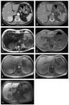
Improved characterization of focal liver lesions with liver-specific gadoxetic acid disodium-enhanced magnetic resonance imaging: a multicenter phase 3 clinical trial
- PMID: 20351497
- PMCID: PMC3036163
- DOI: 10.1097/RCT.0b013e3181c89d87
Improved characterization of focal liver lesions with liver-specific gadoxetic acid disodium-enhanced magnetic resonance imaging: a multicenter phase 3 clinical trial
Abstract
Objectives: To evaluate the safety of gadoxetic acid disodium (Gd-EOB-DTPA) magnetic resonance imaging (MRI) and its efficacy in characterizing liver lesions.
Methods: Lesion characterization and classification using combined (unenhanced and Gd-EOB-DTPA-enhanced) MRI were compared with those using unenhanced MRI and contrast-enhanced spiral computed tomography (CT) using on-site clinical and off-site blinded evaluations for patients with focal liver lesions.
Results: Gadoxetic acid disodium was well tolerated in this study. For the clinical evaluation, more lesions were correctly characterized using combined (unenhanced and Gd-EOB-DTPA-enhanced) MRI than using unenhanced MRI and spiral CT (96% vs 84% and 85%, respectively; P < or = 0.0008). For the blinded evaluation, more lesions were correctly characterized using combined MRI compared with using unenhanced MRI (61%-76% vs 48%-65%, respectively; P < or = 0.0012 for 2/3 readers); when compared with spiral CT, a similar proportion of lesions were correctly characterized.
Conclusions: Gadoxetic acid disodium-enhanced MRI is of clinical benefit relative to unenhanced MRI and spiral CT for a radiological diagnosis of liver lesions.
Figures


Similar articles
-
Detection and characterization of focal liver lesions: a Japanese phase III, multicenter comparison between gadoxetic acid disodium-enhanced magnetic resonance imaging and contrast-enhanced computed tomography predominantly in patients with hepatocellular carcinoma and chronic liver disease.Invest Radiol. 2010 Mar;45(3):133-41. doi: 10.1097/RLI.0b013e3181caea5b. Invest Radiol. 2010. PMID: 20098330 Clinical Trial.
-
Diagnostic efficacy of gadoxetic acid (Primovist)-enhanced MRI and spiral CT for a therapeutic strategy: comparison with intraoperative and histopathologic findings in focal liver lesions.Eur Radiol. 2008 Mar;18(3):457-67. doi: 10.1007/s00330-007-0716-9. Epub 2007 Dec 6. Eur Radiol. 2008. PMID: 18058107
-
Diagnostic performance and description of morphological features of focal nodular hyperplasia in Gd-EOB-DTPA-enhanced liver magnetic resonance imaging: results of a multicenter trial.Invest Radiol. 2008 Jul;43(7):504-11. doi: 10.1097/RLI.0b013e3181705cd1. Invest Radiol. 2008. PMID: 18580333 Clinical Trial.
-
Focal liver lesion detection and characterization with GD-EOB-DTPA.Clin Radiol. 2011 Jul;66(7):673-84. doi: 10.1016/j.crad.2011.01.014. Epub 2011 Apr 23. Clin Radiol. 2011. PMID: 21524416 Review.
-
Diagnostic performance of contrast-enhanced multidetector computed tomography and gadoxetic acid disodium-enhanced magnetic resonance imaging in detecting hepatocellular carcinoma: direct comparison and a meta-analysis.Abdom Radiol (NY). 2016 Oct;41(10):1960-72. doi: 10.1007/s00261-016-0807-7. Abdom Radiol (NY). 2016. PMID: 27318936 Free PMC article.
Cited by
-
Hepatobiliary MR imaging with gadolinium-based contrast agents.J Magn Reson Imaging. 2012 Mar;35(3):492-511. doi: 10.1002/jmri.22833. J Magn Reson Imaging. 2012. PMID: 22334493 Free PMC article. Review.
-
Respiratory motion artefacts in dynamic liver MRI: a comparison using gadoxetate disodium and gadobutrol.Eur Radiol. 2015 Nov;25(11):3207-13. doi: 10.1007/s00330-015-3736-x. Epub 2015 Apr 23. Eur Radiol. 2015. PMID: 25903709
-
Benign liver tumors in pediatric patients - Review with emphasis on imaging features.World J Gastroenterol. 2015 Jul 28;21(28):8541-61. doi: 10.3748/wjg.v21.i28.8541. World J Gastroenterol. 2015. PMID: 26229397 Free PMC article. Review.
-
Update on the Pathology of Pediatric Liver Tumors: A Pictorial Review.Diagnostics (Basel). 2023 Nov 24;13(23):3524. doi: 10.3390/diagnostics13233524. Diagnostics (Basel). 2023. PMID: 38066766 Free PMC article. Review.
-
Safety and Efficacy of Gadoxetate Disodium-Enhanced Liver MRI in Pediatric Patients Aged >2 Months to <18 Years-Results of a Retrospective, Multicenter Study.Magn Reson Insights. 2016 Jul 21;9:21-8. doi: 10.4137/MRI.S39091. eCollection 2016. Magn Reson Insights. 2016. PMID: 27478381 Free PMC article.
References
-
- Hamm B, Thoeni RF, Gould RG, et al. Focal liver lesions: characterization with nonenhanced and dynamic contrast material-enhanced MR imaging. Radiology. 1994;190:417–423. - PubMed
-
- Mitchell DG, Saini S, Weinreb J, et al. Hepatic metastases and cavernous hemangiomas: distinction with standard- and triple-dose gadoteridol-enhanced MR imaging. Radiology. 1994;193:49–57. - PubMed
-
- Petersein J, Spinazzi A, Giovagnoni A, et al. Focal liver lesions: evaluation of the efficacy of gadobenate dimeglumine in MR imaging—a multicenter phase III clinical study. Radiology. 2000;215:727–736. - PubMed
-
- Whitney WS, Herfkens RJ, Jeffrey RB, et al. Dynamic breath-hold multiplanar spoiled gradient-recalled MR imaging with gadolinium enhancement for differentiating hepatic hemangiomas from malignancies at 1.5 T. Radiology. 1993;189:863–870. - PubMed
-
- Federle M, Chezmar J, Rubin DL, et al. Efficacy and safety of mangafodipir trisodium (MnDPDP) injection for hepatic MRI in adults: results of the U.S. Multicenter phase III clinical trials. Efficacy of early imaging. J Magn Reson Imaging. 2000;12:689–701. - PubMed
Publication types
MeSH terms
Substances
Grants and funding
LinkOut - more resources
Full Text Sources
Medical

