. 2010 May;11(5):395-402.
doi: 10.1038/ni.1864. Epub 2010 Mar 28.

The AIM2 inflammasome is essential for host defense against cytosolic bacteria and DNA viruses

Affiliations

The AIM2 inflammasome is essential for host defense against cytosolic bacteria and DNA viruses

Vijay A K Rathinam et al. Nat Immunol. 2010 May.

Abstract

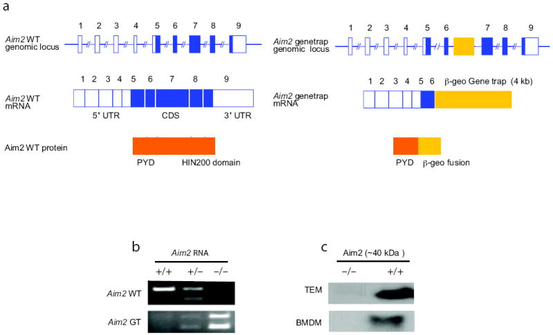
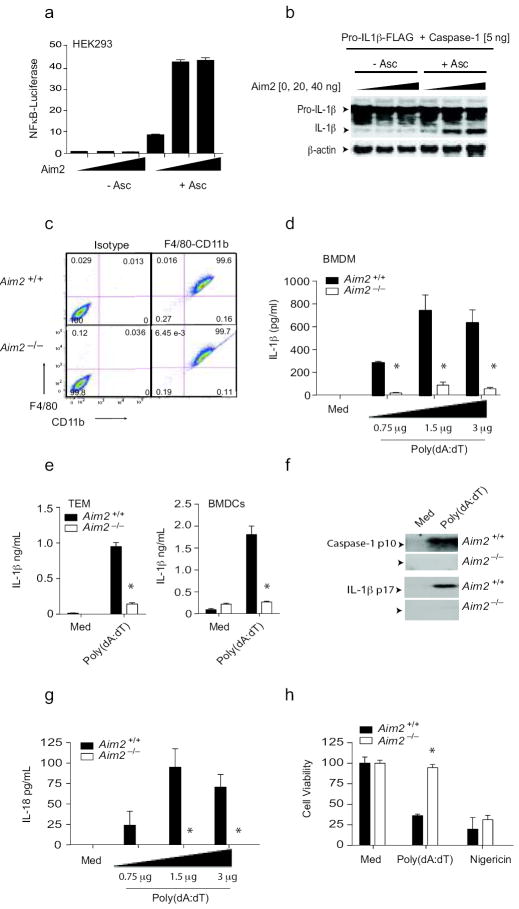
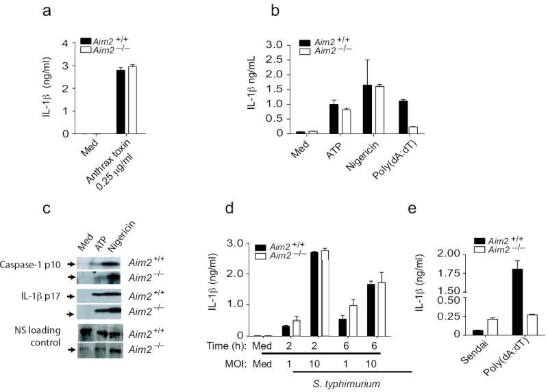
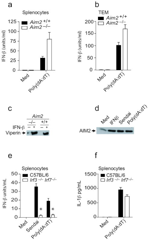
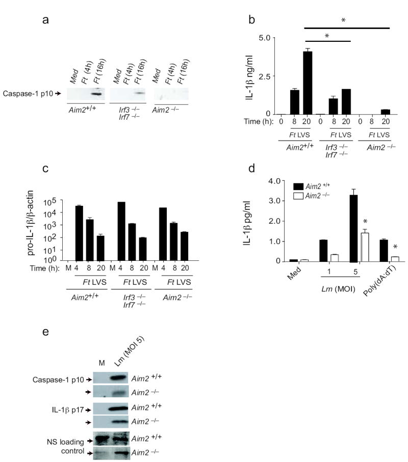
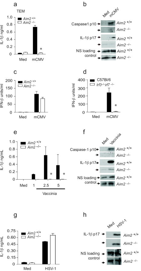
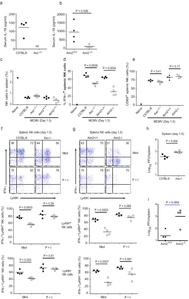
Inflammasomes regulate the activity of caspase-1 and the maturation of interleukin 1beta (IL-1beta) and IL-18. AIM2 has been shown to bind DNA and engage the caspase-1-activating adaptor protein ASC to form a caspase-1-activating inflammasome. Using Aim2-deficient mice, we identify a central role for AIM2 in regulating caspase-1-dependent maturation of IL-1beta and IL-18, as well as pyroptosis, in response to synthetic double-stranded DNA. AIM2 was essential for inflammasome activation in response to Francisella tularensis, vaccinia virus and mouse cytomegalovirus and had a partial role in the sensing of Listeria monocytogenes. Moreover, production of IL-18 and natural killer cell-dependent production of interferon-gamma, events critical in the early control of virus replication, were dependent on AIM2 during mouse cytomegalovirus infection in vivo. Collectively, our observations demonstrate the importance of AIM2 in the sensing of both bacterial and viral pathogens and in triggering innate immunity.


Conflict of interest statement

Competing financial interest. The authors declare no competing financial interest.

Figures

Figure 1
Figure 1
Characterization of Aim2-genetrap mice. a Schematic of wild-type and gene-trapped Aim2 allele. b. RT-PCR analysis shown the absence of wild-type (WT) Aim2 transcripts in the Aim2-genetrap mice. WT Aim2 RNA was detected by primers F and R1 and gene-trapped Aim2 RNA was detected by primers F and R2. c. Immunoblotting of Aim2 protein in peritoneal and bone marrow derived macrophages from Aim2+/+ and Aim2-/- mice. Equal amounts of protein were loaded for all the samples and Aim2 was detected using a rabbit polyclonal antibody.
Figure 2
Figure 2
Aim2 is essential for inflammasome activation by DNA. a. 293T cells were transfected with indicated plasmids together with a NF-κB reporter and TK Renilla-luciferase reporter gene. After 48 h, luciferase activity was measured. b. 293T cells were transfected with the indicated plasmids together with murine pro-IL-1β-Gluc/Flag. After 24 h, cell lysates were immunoblotted for pro-IL1β-Flag, IL-1β-Flag and β-actin. c. F4/80 and CD11b expression by BMDM from Aim2+/+ and Aim2-/- mice. d-f. LPS (200 ng/ml) primed Aim2+/+ and Aim2-/- BMDM, thioglycollate-elicited macrophages (TEM), and BMDCs were transfected with poly(dA-dT) (0.75 – 3 μg/106 cells) for 6 h. The secreted IL-1β (d-e) and the cleaved forms of IL-1β and caspase 1 (f) in the culture supernatants were analyzed. g. Aim2+/+ and Aim2-/- BMDM were treated as above and the IL-18 levels in the supernatants at 6 h were measured by ELISA. (h). Aim2+/+ and Aim2-/- TEM were treated as indicated for 24 h and cell viability was reported as % of viability of medium treated cells. Asterisks indicate P values of < 0.05 for Aim2+/+ versus Aim2-/-. Data are presented as mean ± SD from one experiment representative of 3 experiments.
Figure 3
Figure 3
Aim2-deficient cells respond normally to other inflammasome activating stimuli. a. BMDM from Aim2+/+, Aim2-/-, and C57Bl/6 mice were primed with LPS (200 ng/ml) for 3 h and stimulated with anthrax toxin units PA and LF. Supernatants were harvested after 6 h and analyzed by ELISA for IL-1β production. b-c. LPS primed Aim2+/+ and Aim2-/- TEM were treated with ATP (5 mM) and Nigericin (5 μM) for 1 h and secretion of IL-1β and cleavage of IL-1β and caspase 1 were analyzed. d. Aim2+/+ and Aim2-/- BMDM were primed with LPS (200 ng/ml) for 3 h and infected with S. typhimurium at MOI 1 and MOI 10. Supernatants were harvested at 2 and 6 h post infection and IL-1β levels were analyzed by ELISA. e. BMDCs were treated with Sendai virus (200 HA units/ml) for 18 h and IL-1β concentrations in the supernatant were measured by ELISA. NS; nonspecific band. Asterisks indicate P values of < 0.05 for Aim2+/+ versus Aim2-/-. Data are presented as mean ± SD from one experiment representative of 3 experiments.
Figure 4
Figure 4
Type I interferon production and signaling remain intact in Aim2-deficient cells. A and e-f. Splenocytes from Aim2+/+, Aim2-/-, Irf3-/- Irf7-/- and C57Bl/6 mice were plated at 2 × 10⁵ cells/well in 96-well plates in antibiotic free DMEM and were treated as indicated above. After 24 h, supernatants were collected and analyzed for IFN-β concentrations. b. TEM from Aim2+/+, Aim2-/-, Irf3-/- Irf7-/-, and C57BL/6 mice were treated as indicated above and supernatants were analyzed for IL-1β and IFN-β concentrations 6 h later. c. BMDM from Aim2+/+ and Aim2-/- mice were treated with 1000 units/ml IFNβ for 18 h and the viperin protein level in the lysates was analyzed by immunoblotting. d. TEM from C57Bl/6 mice were treated with 1000 units/ml IFNβ, 200 HA units/ml Sendai virus, and poly(dA-dT)1.5 μg/106 cells for 18 h and the Aim2 protein amounts in lysates was analyzed. Asterisks indicate P values of < 0.05 for Aim2+/+ versus Aim2-/-. Data are presented as mean ± SD from one experiment representative of 3 experiments.
Figure 5
Figure 5
Aim2 mediates inflammasome activation in response to bacterial pathogens. TEM from Aim2+/+, Aim2-/-, Irf3-/- Irf7-/- and C57Bl/6 mice were infected with F. tularensis LVS (MOI = 50) as indicated in the materials and methods. At indicated time points after infection, cleaved caspase 1 (a) and IL-1β (b) in the supernatants and the mRNA levels of pro-IL-1β (c) were measured. d-e. LPS primed Aim2+/+ and Aim2-/- TEM were infected with L. monocytogenes at MOI of 1 and 5 and supernatants were analyzed for secreted IL-1β by ELISA (d) and cleaved caspase 1 and IL-1β by immunoblotting (e). NS; nonspecific band. Asterisks indicate P values of < 0.05 for Aim2+/+ versus Aim2-/-. Data are presented as mean ± SD from one experiment representative of 2 or 3 experiments.
Figure 6
Figure 6
Aim2 is essential for inflammasome activation by DNA viruses. a-b. Aim2-/- and Aim2-/- TEM were primed with LPS for 3 – 4 h and stimulated with mCMV at an MOI of 10 for 20 h. IL-1β concentrations were measured by ELISA and cleaved caspase 1 p10 and IL-1β p17 in the supernatants by immunoblotting. c-d. TEM from Aim2+/+, Aim2-/-, Irf3-/- Irf7-/- and C57Bl/6 mice were stimulated with CMV as indicated above and IFN-β concentrations were measured by ELISA. e-f. Aim2+/+ and Aim2-/- BMDM and BMDCs were infected with vaccinia virus as described for mCMV and the supernatants were analyzed for caspase 1 and IL-1β. g-h. Aim2+/+ and Aim2-/- TEM were LPS primed and treated with HSV-1 at an MOI of 40 and at 18 h post infection, IL-1β concentrations in the supernatants were determined by ELISA and immunoblotting. NS; nonspecific band. Asterisks indicate P values of < 0.05 for Aim2+/+ versus Aim2-/-. Data are presented as mean ± SD from one experiment representative of 3 experiments.
Figure 7
Figure 7
Aim2- and Asc-dependent IL-18 production and NK cell IFN-γ production. a-b. Serum IL-18 concentrations in C57BL/6, Asc-/-, Aim2+/+ and Aim2-/- mice infected with MCMV. c Proportions of NK1.1+ NKp46+ CD3- NK cells in the spleens of infected mice as well as an uninfected C57BL/6 control. d and e At 36 h p.i., splenocytes were cultured ex vivo in medium containing brefeldin A for 4 h with no additional stimuli. Intracellular IFN-γ (d) and surface CD69 (e) expressions were determined on total splenic NK cells. f-g. Splenocytes cultured in either medium without additional stimuli or medium containing PMA (50 ng/mL) and ionomycin (500 ng/mL) (denoted P + I) were stained for surface Ly49H and intracellular IFN-γ. Representative staining of gated NK cells is shown for (f) C57BL/6 and Asc-/- as well as (g) Aim2+/+ and Aim2-/- mice. Numbers in upper quadrants are proportion of Ly49H+ or Ly49H- NK cells that are IFN-γ+. Proportions of Ly49H+ and Ly49H- NK cells expressing IFN-γ are shown for individual mice in lower plots. In lower panels, black squares are C57BL/6 mice and white squares are Asc-/-, while black circles are Aim2+/+ and white circles are Aim2-/- (h-i) Viral titers in the spleen at 36 h p.i. nd; not detectable. P values were determined by unpaired two-tailed Student’s T test. Data are from one experiment representative of 2 experiments.

Comment in

Similar articles

Cited by

References

    1. Kawai T, Akira S. Toll-like receptor and RIG-I-like receptor signaling. Ann N Y Acad Sci. 2008;1143:1–20. - PubMed
    1. Takeda K, Akira S. Toll-like receptors in innate immunity. Int Immunol. 2005;17:1–14. - PubMed
    1. Huysamen C, Brown GD. The fungal pattern recognition receptor, Dectin-1, and the associated cluster of C-type lectin-like receptors. FEMS Microbiol Lett. 2009;290:121–128. - PMC - PubMed
    1. Yoneyama M, Fujita T. RIG-I family RNA helicases: cytoplasmic sensor for antiviral innate immunity. Cytokine Growth Factor Rev. 2007;18:545–551. - PubMed
    1. Martinon F, Mayor A, Tschopp J. The inflammasomes: guardians of the body. Annu Rev Immunol. 2009;27:229–265. - PubMed

Publication types

MeSH terms