. 2010 May;11(5):385-93.
doi: 10.1038/ni.1859. Epub 2010 Mar 28.

The AIM2 inflammasome is critical for innate immunity to Francisella tularensis

Affiliations

The AIM2 inflammasome is critical for innate immunity to Francisella tularensis

Teresa Fernandes-Alnemri et al. Nat Immunol. 2010 May.

Abstract

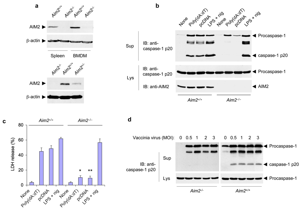
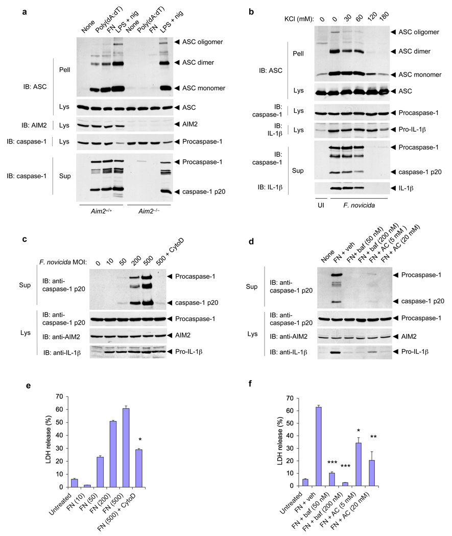
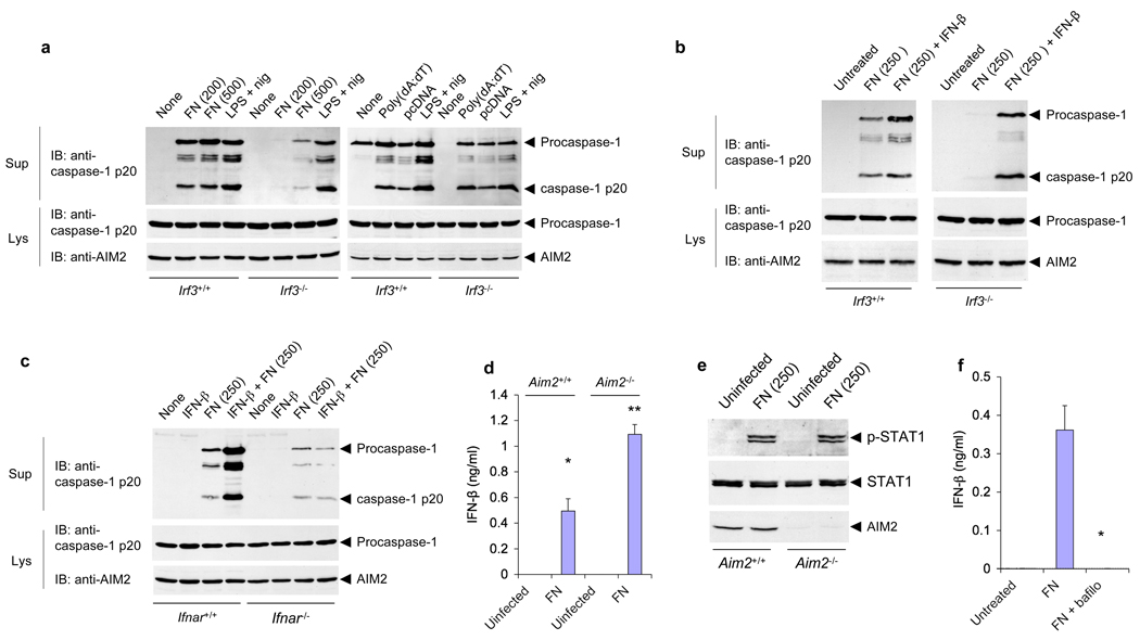
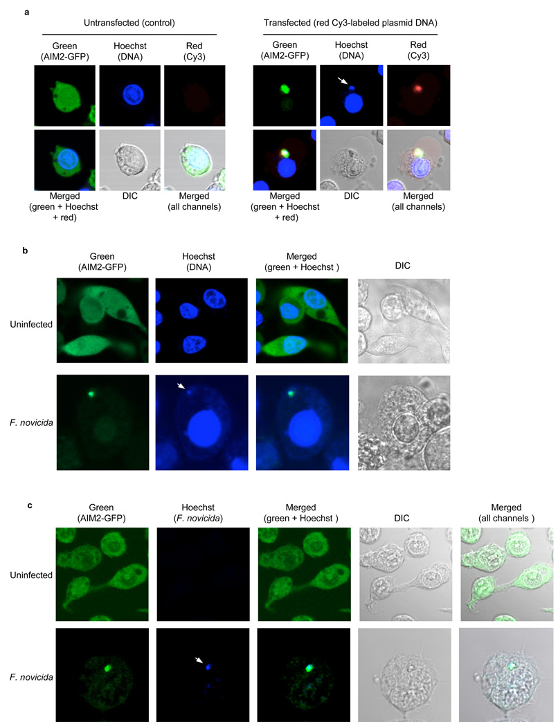
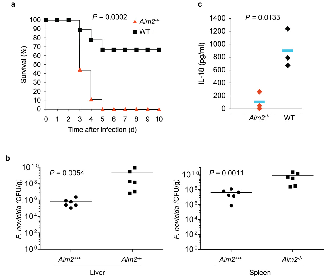
Francisella tularensis, the causative agent of tularemia, infects host macrophages, which triggers production of the proinflammatory cytokines interleukin 1beta (IL-1beta) and IL-18. We elucidate here how host macrophages recognize F. tularensis and elicit this proinflammatory response. Using mice deficient in the DNA-sensing inflammasome component AIM2, we demonstrate here that AIM2 is required for sensing F. tularensis. AIM2-deficient mice were extremely susceptible to F. tularensis infection, with greater mortality and bacterial burden than that of wild-type mice. Caspase-1 activation, IL-1beta secretion and cell death were absent in Aim2(-/-) macrophages in response to F. tularensis infection or the presence of cytoplasmic DNA. Our study identifies AIM2 as a crucial sensor of F. tularensis infection and provides genetic proof of its critical role in host innate immunity to intracellular pathogens.


Figures

Figure 1
Figure 1
Disruption of mouse Aim2 abolishes activation of the inflammasome by cytoplasmic DNA and vaccinia virus. (a) Immunoblot analysis of the expression of AIM2 in spleens and BMDMs from Aim2+/+ and Aim2−/− littermates (top) and in spleens from Aim2−/−, Aim2+/+ and Aim2+/− mice (bottom), assessed with an antibody specific for mouse AIM2. β-actin serves as a loading control. (b) Immunoblot (IB) analysis of mouse procaspase-1, caspase-1 (p20 subunit) and/or AIM2 in culture supernatants (Sup) and lysates (Lys) of Aim2+/+ and Aim2−/− BMDMs left untreated (None) or transfected with the synthetic DNA poly(dA:dT) or plasmid DNA (pcDNA), or treated for 5 h with LPS (500 ng/ml) followed by nigericin (2.5 µM) for 45 min (LPS + nig), assessed with monoclonal antibody to mouse caspase-1 p20 (anti-p20). (c) Release of LDH into culture supernatants of the BMDMs described in b, presented relative to the total cellular LDH content. *P < 0.05 and **P < 0.01, Aim2+/+ versus Aim2−/− (Student’s t-test). (d) Immunoblot analysis of mouse procaspase-1 and caspase-1 in culture supernatants and lysates of mouse Aim2−/− and Aim2+/+ BMDMs infected for 18 h with vaccinia virus (multiplicity of infection (MOI), above lanes). Data are representative of at least three experiments (mean and s.d. in c).
Figure 2
Figure 2
AIM2 is required for F. novicida–induced activation of the inflammasome. (a) Immunoblot analysis of mouse procaspase-1, caspase-1, IL-1β, AIM2 and/or pro-IL-1β in culture supernatants and lysates of mouse Aim2−/− and Aim2+/+ macrophages left untreated or infected for 6 h with F. novicida (FN; MOI in parentheses above lanes) or treated with LPS and nigericin as described in Figure 1b. (b) Release of LDH into culture supernatants of the macrophages in a. *P < 0.05, **P < 0.01 and ***P < 0.005, Aim2+/+ versus Aim2−/− (Student’s t-test). (c) Confocal live-cell microsopy of Aim2−/− and Aim2+/+ BMDMs left uninfected (UI) or infected for 6 h with F. novicida; nuclei were stained with Hoechst stain (blue). Images are merged differential interference contrast and Hoechst channels. Original magnification, x40. (d) Immunoblot analysis of mouse procaspase-1, caspase-1, ASC, AIM2 and/or Nlrp3 in culture supernatants and lysates of mouse wild-type, ASC-deficient (Pycard−/−; called ‘Asc−/−’ here) and Nlrp3−/− macrophages infected with F. novicida for 6 h or for 24 h (far right; MOI in parentheses above lanes). (e) Immunoblot analysis of mouse procaspase-1, caspase-1, IL-1β, pyrin and/or pro-IL-1β in culture supernatants and lysates of mouse pyrin-deficient (Mefv−/−) and pyrin-sufficient (Mefv+/+) macrophages infected for 6 h with F. novicida (MOI in parentheses above lanes) or treated with LPS and nigericin as described in a. (f) Release of LDH into culture supernatants of the macrophages in e. Data are representative of at least three experiments (mean and s.d. in b,f).
Figure 3
Figure 3
Role of the ASC pyroptosome, potassium depletion, actin polymerization and lysosomal acidification in activation of the AIM2 inflammasome by F. novicida. (a) Immunoblot analysis of ASC pyroptosomes in Aim2+/+ and Aim2−/− BMDMs transfected with poly(dA:dT), infected for 6 h with F. novicida (MOI, 500), or treated with LPS and nigericin as described in Figure 1b; pellets of whole-cell lysates centrifuged at 3,800g followed by crosslinking with disuccinimidyl suberate (Pell), as well as cell lysates and culture supernatants, were hybridized with anti–mouse ASC, anti–mouse AIM2 and anti-caspase-1. (b) Immunoblot analysis of ASC pyroptosomes in Aim2+/+ BMDMs left uninfected or infected for 6 h with F. novicida (MOI, 500) in the presence of increasing concentrations of KCl in the culture medium, then fractionated and analyzed as in a. (c,d) Immunoblot analysis of mouse procaspase-1, caspase-1, AIM2 and/or pro-IL-1β in culture supernatants and lysates of Aim2+/+ BMDMs infected for 6 h with F. novicida (MOI, above lanes) in the presence (+ cytoD) or absence of cytochalasin D (c) or infected for 6 h with the F. novicida (MOI, 250) in the presence of vehicle (+ veh), bafilomycin (+ baf) or NH4Cl (+ AC; d). (e,f) Release of LDH into culture supernatants of the BMDMs in c (e) and d (f). (e) *P < 0.001, with versus without cytochalasin D (Student’s t-test); (f) *P < 0.05, **P < 0.01 and ***P < 0.001, F. novicida versus vehicle (Student’s t-test). Data are representative of two (a–d) or three (e,f) experiments (mean and s.d. in e,f).
Figure 4
Figure 4
IRF3 signaling is required for activation of the AIM2 inflammasome by F. novicida but not by liposome-delivered DNA. (a,b) Immunoblot analysis of mouse procaspase-1, caspase-1 and/or AIM2 in culture supernatants and lysates of mouse Irf3−/− and Irf3+/+ macrophages infected for 6 h with F. novicida (MOI, in parentheses above lanes), treated with LPS and nigericin as described in Figure 1b, or transfected with poly(dA:dT) (a), or infected with F. novicida (MOI, 250) in the presence or absence of IFN-β (b). (c) Immunoblot analysis of mouse procaspase-1, caspase-1 and/or AIM2 in culture supernatants and lysates of Ifnar1−/− and Ifnar1+/+ macrophages left untreated or treated for 2 h with IFN-β alone or followed by infection for 6 h with F. novicida (MOI, in parentheses above lanes). (d) Enzyme-linked immunosorbent assay of IFN-β in culture supernatants of Aim2−/− and Aim2+/+ macrophages left uninfected or infected for 6 h with F. novicida (MOI, 250). *P < 0.05 and **P < 0.005 (Student’s t-test). (e) Immunoblot analysis of mouse STAT1 phosphorylated at Tyr701 (p-STAT1), total STAT1 and AIM2 in lysates of mouse Aim2−/− and Aim2+/+ macrophages left uninfected or infected with F. novicida (MOI, 250). (f) Enzyme-linked immunosorbent assay of IFN-β in culture supernatants of Aim2+/+ macrophages left untreated or infected for 6 h with F. novicida (MOI, 250) in the presence (+ bafilo) or absence of bafilomycin (50 nM). *P < 0.01 (Student’s t-test). Data are representative of two (a,c) or three (b,d–f) experiments (mean and s.d. in d,f).
Figure 5
Figure 5
Cytoplasmic DNA secreted by Francisella induces AIM2 oligomerization. (a), Enlarged confocal live cell images of Nlrp3−/−-AIM2-EGFP-N1 BMDM following transfection with Cy-3™-labeled DNA (right panels) or nothing (control, left panels). The white arrow in the blue channel (right panels) indicates staining of the clustered cytoplasmic DNA with the blue Hoechst stain, which specifically stains DNA. (b), Enlarged confocal live cell images of Nlrp3−/−-AIM2-EGFP-N1 BMDM left uninfected (upper panels) or infected with F. novicida (lower panels) for 6 h and then stained with Hoechst stain before microscopy. The white arrow in the blue channel (lower panels) indicates staining of the AIM2-GFP cluster with the DNA-specific blue Hoechst stain. (c), Enlarged confocal cell images of Nlrp3−/−-AIM2-EGFP-N1 BMDM left uninfected (upper panels) or infected with Hoechst-labeled F. novicida (lower panels) for 6 h and then fixed on coverslips before confocal microscopy. The white arrow in the blue channel (lower panels) indicates the Hoechst-labeled Francisella cytoplasmic DNA. Additional representative confocal images of Nlrp3−/−-AIM2-EGFP-N1 BMDM infected with unstained or Hoechst-pre-stained F. novicida are shown in supplementary Figure 11. DIC, differential interference contrast. Original magnification, x40. Data are representative of at least three (a,b) or two (c) experiments.
Figure 6
Figure 6
The AIM2 inflammasome is critical for innate immunity against Francisella infection. (a), Survival of Aim2+/+ and Aim2−/− mice injected subcutaneously with F. novicida (1.5 × 10⁵ CFU) (Aim2+/+ n = 9, Aim2−/− n=9), and monitored over a period of 3 weeks. 66% of Aim2+/+ survived beyond 3 weeks post-infection. (b), Livers and spleens were harvested 48 h post-infection of mice subcutaneously with F. novicida (1.0 × 10⁵ CFU) homogenized, and dilutions plated on Cystine Heart Agar plates for enumeration of CFU. Bacterial counts from the livers and spleens of the Aim2−/− were significantly higher compared with Aim2+/+ mice (P 0.0054, and 0.0011, respectively). (c) Enzyme-linked immunosorbent assay of IL-18 in serum from Aim2+/+ mice (n = 3) and Aim2−/− mice (n = 3) at 1 d after subcutaneous infection with F. novicida. In b,c, each symbol represents an individual mouse; small horizontal lines indicate the mean. P values, Kaplan-Meier log-rank test (a) or Student’s t-test (b,c). Data are representative of two experiments.

Comment in

Similar articles

Cited by

References

    1. Akira S, Uematsu S, Takeuchi O. Pathogen recognition and innate immunity. Cell. 2006;124:783–801. - PubMed
    1. Kumar H, Kawai T, Akira S. Pathogen recognition in the innate immune response. Biochem J. 2009;420:1–16. - PubMed
    1. Fernandes-Alnemri T, Yu JW, Datta P, Wu J, Alnemri ES. AIM2 activates the inflammasome and cell death in response to cytoplasmic DNA. Nature. 2009;458:509–513. - PMC - PubMed
    1. Hornung V, et al. AIM2 recognizes cytosolic dsDNA and forms a caspase-1-activating inflammasome with ASC. Nature. 2009;458:514–518. - PMC - PubMed
    1. Roberts TL, et al. HIN-200 proteins regulate caspase activation in response to foreign cytoplasmic DNA. Science. 2009;323:1057–1060. - PubMed

Publication types

MeSH terms