

Primary laryngeal tuberculosis mimicking laryngeal carcinoma: CT scan features
- PMID: 20351985
- PMCID: PMC2844739
- DOI: 10.4103/0971-3026.59745
Primary laryngeal tuberculosis mimicking laryngeal carcinoma: CT scan features
Abstract
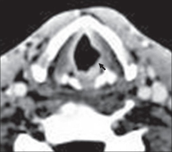
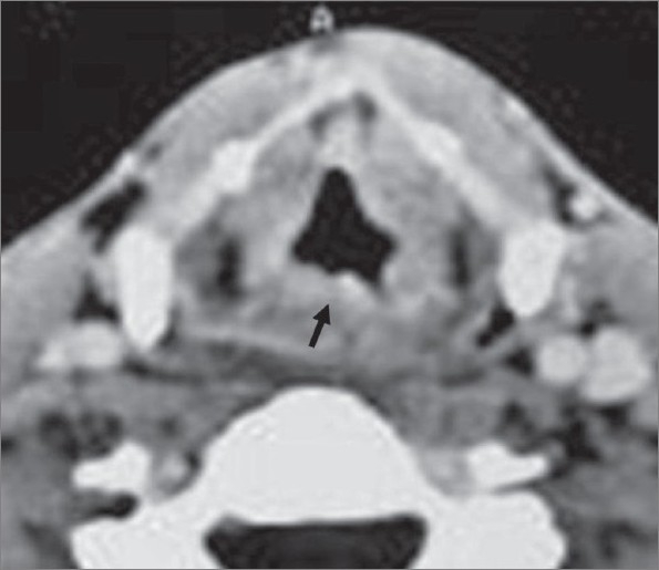
Laryngeal tuberculosis is a rare disease. It is almost always associated with pulmonary tuberculosis. It occurs generally in adults without BCG vaccination or in cases of the acquired immune deficiency syndrome. On laryngoscopy and imaging, it often simulates laryngeal carcinoma, and confirmation is always histological. We report the case of a 36-year-old man who presented to our hospital with dysphonia and dysphagia. Laryngoscopy revealed a lesion of the left vocal cord and the ventricular strip. CT scan found focal, regular thickening of the left vocal cord, associated with irregular thickening of the posterior laryngeal wall. A biopsy confirmed the diagnosis of tuberculosis.
Keywords: Carcinoma; larynx; tuberculosis.
Conflict of interest statement
Figures
References
-
- Kozakiewicz J, Dec M, Gorczyca-Tarnowska J. The rare case of primary isolated tuberculosis in a 19 year-old patient. Otolaryngol Pol. 2006;60:607–9. - PubMed
-
- Loehrl TA, Smith TL. Inflammatory and granulomatous lesions of the larynx and pharynx. Am J Med. 2001;111:113–7. - PubMed
-
- Kouassi B, Ette A, Bamba M, Haffner G, Fakhry K, Ehouo F. A propos de 2 cas de tuberculose laryngée primitive. Les Cahiers d'ORL. 1984;19:841–6.
-
- Aspestrand F, Kolbenstvedt A, Boysen M. CT findings in benign expansions of the larynx. J Comput Assist Tomogr. 1989;13:222–5. - PubMed
LinkOut - more resources
Full Text Sources