

doi: 10.4103/0971-3026.59757.
CT mimics of peritoneal carcinomatosis
Affiliations
- PMID: 20351997
- PMCID: PMC2844752
- DOI: 10.4103/0971-3026.59757
Item in Clipboard
CT mimics of peritoneal carcinomatosis
Indian J Radiol Imaging.
2010 Feb.
Abstract
Peritoneal carcinomatosis is a term used to describe widespread metastases of cancerous tumors in the peritoneal cavity. It is most common in carcinomas of the gastrointestinal tract (GIT) and ovaries, and must be considered to be the main diagnosis even when the primary is not known. A wide variety of disease processes mimic peritoneal carcinomatosis. Precise diagnosis based on imaging alone is often difficult and very often the final diagnosis is only obtained after appropriate histopathology or microbiology.
Keywords: Carcinomatosis; neoplastic; peritoneal.
Conflict of interest statement
Figures

Peritoneal carcinomatosis. Omental caking (arrow) and ascites (arrowhead) are seen on an axial, contrast-enhanced CT scan of the abdomen, in a 55-year-old woman, a known case of carcinoma ovary with raised CA-125 levels

Peritoneal carcinomatosis. Axial contrast-enhanced CT scan of the mid-pelvis (a) shows a heterogeneously enhancing peritoneal/omental deposit (arrow). An axial image (b), a little inferior to A shows a large heterogeneous mass (arrow) in the pelvis with solid and cystic components. Biopsy showed papillary adenocarcinoma of the ovary

Lymphoma. A 36-year-old male presented with abdominal pain. Axial, contrast-enhanced CT scans of the abdomen show diffuse omental thickening (arrow in a) with bowel wall thickening in the ascending colon (arrow in b) and small bowel (arrow in c). Biopsy was consistent with small-cell, cleaved non-Hodgkin lymphoma

Lymphoma. A 45-year-old male with non-Hodgkin lymphoma has para-aortic and mesenteric lymphadenopathy (arrows) along with splenomegaly (arrowhead), on a contrast-enhanced, axial CT scan of the abdomen

Lymphoma. A 34-year-man with 1-month history of abdominal pain has omental thickening (arrow in a) and an epigastric mass (arrow in b), on contrast-enhanced, axial CT scans of the abdomen. Histopathology was suggestive of non-Hodgkin lymphoma

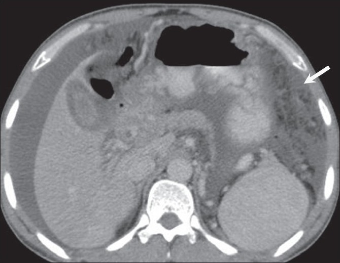
Peritoneal mesothelioma. A 60-year-old woman presented with abdominal distension and loss of appetite. Axial, contrastenhanced images of the abdomen show a lobulated heterogenously enhancing intraperitoneal lesion in the left hemi-abdomen (arrow in a) with ascites (arrow in b); however, no calcification was seen within the mass. Biopsy was suggestive of primary peritoneal mesothelioma

GIST. Axial, contrast-enhanced CT scan of the abdomen in a 65-year-old male with diffuse abdominal pain shows a large, heterogenously enhancing intraperitoneal mass having central necrosis (arrow in a) with multiple peritoneal deposits (arrow in b). This was confirmed on histopathology

Tuberculosis. Axial, contrast-enhanced CT scan in a 35-year-old man with abdominal tuberculosis shows diffuse omental thickening (arrow)
Tuberculosis. Axial, contrast-enhanced CT scan in a 40- year- old woman with abdominal pain shows omental thickening (arrow) along with ascites. A possibility of peritoneal carcinomatosis was considered. Histopathology was suggestive of tuberculosis
References
-
- Pickhardt PJ, Bhalla S. Primary neoplasms of peritoneal and sub-peritoneal origin: CT Findings - Radiographics. 2005;25:983–95. - PubMed
-
- Balachandran A, Silverman PM. Mesenteric and Omental lesions. In: Gore, Levine, editors. Textbook of Gastrointestinal radiology. 3rd ed. vol. 2. Saunders; 2143 and 2139.
-
- Gollub MJ. Imaging of gastrointestinal lymphoma. Radiol Clin North Am. 2008;46:287–312. - PubMed
-
- Horger M, Müller-Schimpfle M, Yirkin I, Wehrmann M, Claussen CD. Extensive peritoneal and omental lymphomatosis with raised CA 125 mimicking carcinomatosis: CT and intraoperative findings. Br J Radiol. 2004;77:71–3. - PubMed
-
- Hardy SM. The Sandwich sign. Radiology. 2003;226:651–2. - PubMed
LinkOut - more resources
Full Text Sources