Paradoxical hepatic tumor: Undifferentiated embryonal sarcoma of the liver
- PMID: 20352000
- PMCID: PMC2844756
- DOI: 10.4103/0971-3026.59760
Paradoxical hepatic tumor: Undifferentiated embryonal sarcoma of the liver
Abstract
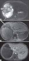
Undifferentiated embryonal sarcoma (UES) is a rare primary malignant tumor of the liver that typically presents in late childhood. We report a case of primary UES, which had a typical paradoxical appearance on different imaging modalities.
Keywords: Liver; children; embryonal sarcoma; imaging; tumor.
Conflict of interest statement
Figures


References
-
- Joshi SW, Merchant NH, Jambhekar NA. Primary multilocular cystic undifferentiated (embryonal) sarcoma of the liver in childhood resembling hydatid cyst of the liver. Br J Radiol. 1997;70:314–6. - PubMed
-
- Wei ZG, Tang LF, Chen ZM, Tang HF, Li MJ. Childhood undifferentiated embryonal liver sarcoma: Clinical features and immunohistochemistry analysis. J Pediatr Surg. 2008;43:1912–9. - PubMed
-
- Buetow PC, Buck JL, Pantongrag-Brown L, Marshall WH, Ros PR, Levine MS, et al. Undifferentiated (embryonal) sarcoma of the liver: Pathologic basis of Imaging findings in 28 cases. Radiology. 1997;203:779–83. - PubMed
-
- Mortelé KJ, Ros PR. Cystic focal liver lesions in the adult: Differential CT and MR imaging features. Radiographics. 2001;21:895–910. - PubMed
-
- Roebuck DJ, Yang WT, Lam WW, Stanley P. Hepatobiliary rhabdomyosarcoma in children: Diagnostic radiology. Pediatr Radiol. 1998;28:101–8. - PubMed
Publication types
LinkOut - more resources
Full Text Sources

