

Persistent mullerian duct syndrome
- PMID: 20352001
- PMCID: PMC2844757
- DOI: 10.4103/0971-3026.59761
Persistent mullerian duct syndrome
Abstract
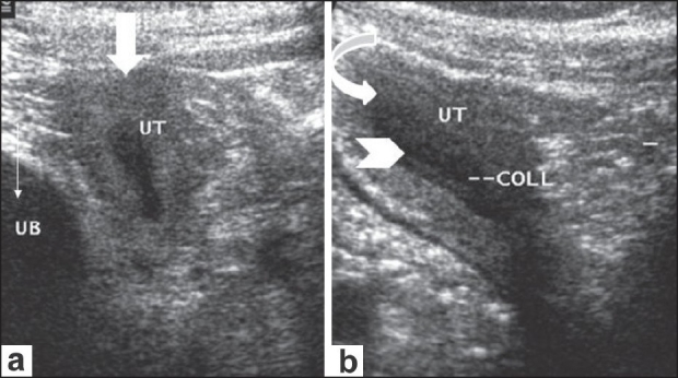
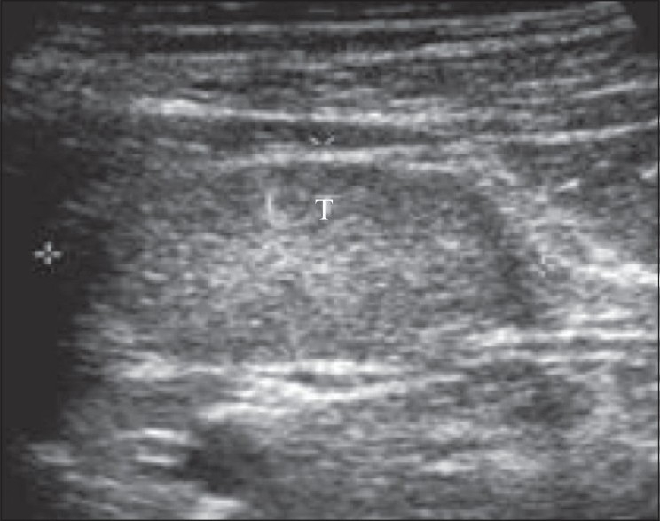
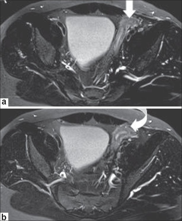
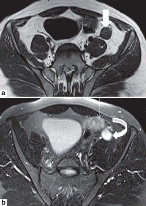
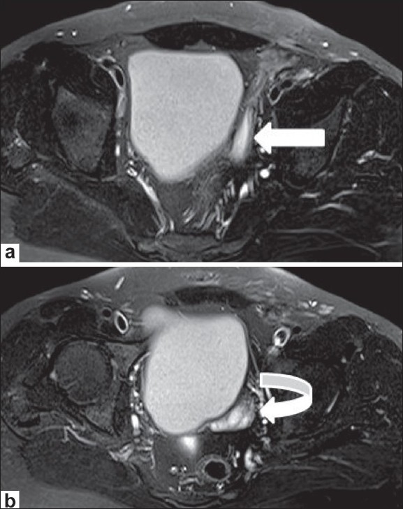
Persistent Mullerian duct syndrome (PMDS) is a rare form of internal male pseudohermaphroditism in which Mullerian duct derivatives are seen in a male patient. This syndrome is characterized by the persistence of Mullerian duct derivatives (i.e. uterus, cervix, fallopian tubes and upper two thirds of vagina) in a phenotypically and karyotypically male patient. In this article we present the USG and MRI features of a case of PMDS with bilateral cryptorchidism and left sided inguinal hernia, containing the uterus and fallopian tubes.
Keywords: Cryptorchidism; MRI; USG; inguinal hernia; mullerian duct derivatives; mullerian inhibiting factor; pseudohermaphroditism.
Conflict of interest statement
Figures
References
-
- Yuksel B, Saygun O, Hengirmen S. Persistent müllerian duct syndrome associated with irreducible inguinal hernia, bilateral cryptorchidism and testicular neoplasia: A case report. Acta Chir Belg. 2006;106:119–20. - PubMed
-
- Gutte AA, Pendharkar PS, Sorte SZ. Transverse testicular ectopia associated with persistent Mullerian duct syndrome – the role of imaging. Br J Radiol. 2008;81:E176–8. - PubMed
-
- Dekker HM, de Jong IJ, Sanders J, Wolf RF. Persistent mullerian duct syndrome. Radiographics. 2003;23:309–13. - PubMed
-
- Clemente A, Macchi V, Berretta M, Morra A. Female form of persistent müllerian duct syndrome: MDCT findings. Clin Imaging. 2008;32:314–7. - PubMed
-
- Wu HC, Chen JH, Lu HF, Shen WC. Persistent mullerian duct syndrome with seminoma. AJR Am J Roentgenol. 2000;174:102–4. - PubMed
Publication types
LinkOut - more resources
Full Text Sources