

Molecular Mechanisms of Neurodegenerative Diseases Induced by Human Retroviruses: A Review
- PMID: 20352020
- PMCID: PMC2845477
- DOI: 10.3844/ajidsp.2009.231.258
Molecular Mechanisms of Neurodegenerative Diseases Induced by Human Retroviruses: A Review
Abstract
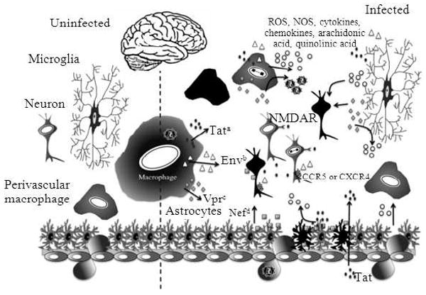
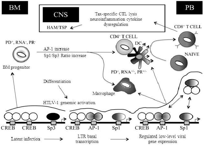
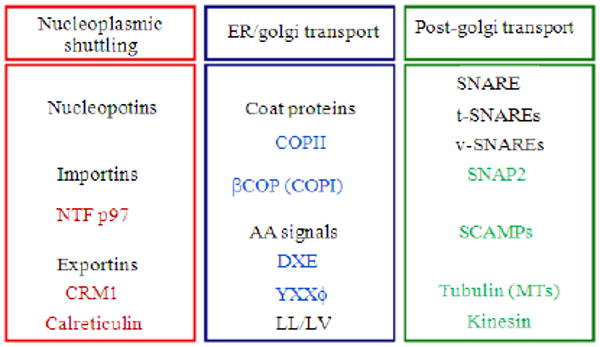
PROBLEM STATEMENT: Infection with retroviruses such as human immunodeficiency virus type 1 (HIV-1) and human T cell leukemia virus type 1 (HTLV-1) have been shown to lead to neurodegenerative diseases such as HIV-associated dementia (HAD) or neuroAIDS and HTLV-1-Associated Myelopathy/Tropical Spastic Paraparesis (HAM/TSP), respectively. APPROACH: HIV-1-induced neurologic disease is associated with an influx of HIV-infected monocytic cells across the blood-brain barrier. Following neuroinvasion, HIV-1 and viral proteins, in addition to cellular mediators released from infected and uninfected cells participate in astrocytic and neuronal dysregulation, leading to mild to severe neurocognitive disorders. RESULTS: The molecular architecture of viral regulatory components including the Long Terminal Repeat (LTR), genes encoding the viral proteins Tat, Vpr and Nef as well as the envelope gene encoding gp120 and gp41 have been implicated in 'indirect' mechanisms of neuronal injury, mechanisms which are likely responsible for the majority of CNS damage induced by HIV-1 infection. The neuropathogenesis of HAM/TSP is linked, in part, with both intra-and extracellular effectors functions of the viral transactivator protein Tax and likely other viral proteins. Tax is traditionally known to localize in the nucleus of infected cells serving as a regulator of both viral and cellular gene expression. CONCLUSION/RECOMMENDATIONS: However, recent evidence has suggested that Tax may also accumulate in the cytoplasm and be released from the infected cell through regulated cellular secretion processes. Once in the extracellular environment, Tax may cause functional alterations in cells of the peripheral blood, lymphoid organs and the central nervous system. These extracellular biological activities of Tax are likely very relevant to the neuropathogenesis of HTLV-1 and represent attractive targets for therapeutic intervention.
Figures
Similar articles
-
Tax secretion from peripheral blood mononuclear cells and Tax detection in plasma of patients with human T-lymphotropic virus-type 1-associated myelopathy/tropical spastic paraparesis and asymptomatic carriers.J Med Virol. 2016 Mar;88(3):521-31. doi: 10.1002/jmv.24342. Epub 2015 Oct 19. J Med Virol. 2016. PMID: 26241614
-
Tropical spastic paraparesis and HTLV-1 associated myelopathy: clinical, epidemiological, virological and therapeutic aspects.Rev Neurol (Paris). 2012 Mar;168(3):257-69. doi: 10.1016/j.neurol.2011.12.006. Epub 2012 Mar 7. Rev Neurol (Paris). 2012. PMID: 22405461 Review.
-
Human T-Lymphotropic Virus Type 1-Induced Overexpression of Activated Leukocyte Cell Adhesion Molecule (ALCAM) Facilitates Trafficking of Infected Lymphocytes through the Blood-Brain Barrier.J Virol. 2016 Jul 27;90(16):7303-7312. doi: 10.1128/JVI.00539-16. Print 2016 Aug 15. J Virol. 2016. PMID: 27252538 Free PMC article.
-
Neuronal molecular mimicry in immune-mediated neurologic disease.Ann Neurol. 1998 Jul;44(1):87-98. doi: 10.1002/ana.410440115. Ann Neurol. 1998. PMID: 9667596
-
Human T cell lymphotropic virus type I genomic expression and impact on intracellular signaling pathways during neurodegenerative disease and leukemia.Front Biosci. 2000 Jan 1;5:D138-68. doi: 10.2741/yao. Front Biosci. 2000. PMID: 10702386 Review.
Cited by
-
Retroviral b-Zip protein (HBZ) contributes to the release of soluble and exosomal immune checkpoint molecules in the context of neuroinflammation.J Extracell Biol. 2023 Jul;2(7):e102. doi: 10.1002/jex2.102. Epub 2023 Jul 17. J Extracell Biol. 2023. PMID: 37547182 Free PMC article.
-
Novel PI3K/Akt inhibitors screened by the cytoprotective function of human immunodeficiency virus type 1 Tat.PLoS One. 2011;6(7):e21781. doi: 10.1371/journal.pone.0021781. Epub 2011 Jul 12. PLoS One. 2011. PMID: 21765914 Free PMC article.
-
Human immunodeficiency virus encephalitis.Indian J Sex Transm Dis AIDS. 2020 Jan-Jun;41(1):108-110. doi: 10.4103/ijstd.IJSTD_112_16. Epub 2020 Jun 18. Indian J Sex Transm Dis AIDS. 2020. PMID: 33062995 Free PMC article.
-
Phosphoglycerate Mutase 1 Promotes Cell Proliferation and Neuroblast Differentiation in the Dentate Gyrus by Facilitating the Phosphorylation of cAMP Response Element-Binding Protein.Neurochem Res. 2019 Feb;44(2):323-332. doi: 10.1007/s11064-018-2678-5. Epub 2018 Nov 20. Neurochem Res. 2019. PMID: 30460638
-
Intracerebroventricular administration of HIV-1 Tat induces brain cytokine and indoleamine 2,3-dioxygenase expression: a possible mechanism for AIDS comorbid depression.Brain Behav Immun. 2011 Nov;25(8):1569-75. doi: 10.1016/j.bbi.2011.05.006. Epub 2011 May 17. Brain Behav Immun. 2011. PMID: 21620953 Free PMC article.
References
-
- Chen W, Tang Z, Fortina P, Patel P, Addya S, Surrey S, Acheampong EA, Mukhtar M, Pomerantz RJ. Ethanol potentiates HIV-1 gp120-induced apoptosis in human neurons via both the death receptor and NMDA receptor pathways. Virology. 2005. pp. 59–73. http://www.ncbi.nlm.nih.gov/pubmed/15749123. - PubMed
-
- McArthur JC. Neurologic manifestations of AIDS. Medicine (Baltimore) 1987. pp. 407–437. http://www.be-md.ncbi.nlm.nih.gov/pubmed/3316921. - PubMed
-
- Silvers JM, Aksenova MV, Aksenov MY, Mactutus CF, Booze RM. Neurotoxicity of HIV-1 Tat protein: Involvement of D1 dopamine receptor. Neurotoxicology. 2007. pp. 1184–1190. http://www.ncbi.nlm.nih.gov/pubmed/17764744. - PMC - PubMed
-
- Childs EA, Lyles RH, Selnes OA, Chen B, Miller EN, et al. Plasma viral load and CD4 lymphocytes predict HIV-associated dementia and sensory neuropathy. Neurology. 1999. pp. 607–613. http://www.neurology.org/cgi/content/abstract/52/3/607. - PubMed
-
- Gendelman HE, Persidsky Y. Infections of the nervous system. Lancet Neurol. 2005. pp. 12–13. http://linkinghub.elsevier.com/retrieve/pii/S1474442204009512. - PMC - PubMed
Grants and funding
LinkOut - more resources
Full Text Sources
Miscellaneous