

Identification of crystallin modifications in the human lens cortex and nucleus using laser capture microdissection and CyDye labeling
- PMID: 20352024
- PMCID: PMC2845665
Identification of crystallin modifications in the human lens cortex and nucleus using laser capture microdissection and CyDye labeling
Abstract
Purpose: With aging, lens crystallins undergo post-translational modifications (PTMs) and these modifications are believed to play a major role in age-related cataract development. The purpose of the present study was to determine the protein profiles of crystallins and their PTMs in the cortical and nuclear regions within an aging human lens to gain a better understanding about changes in crystallins as fiber cells migrate from cortical to nuclear region.
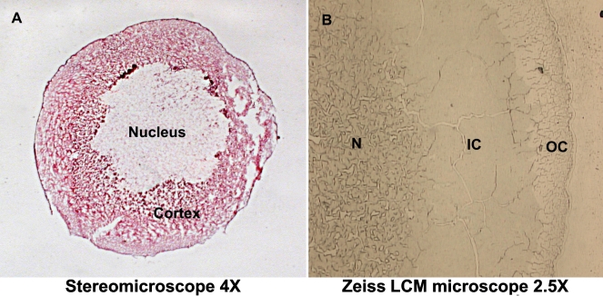
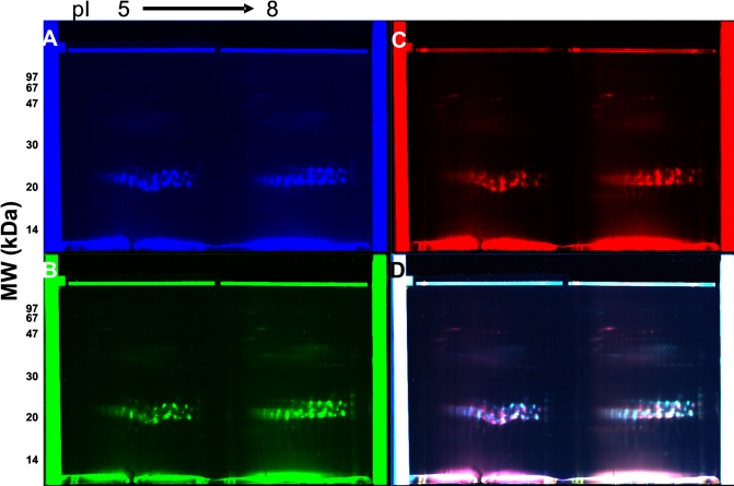
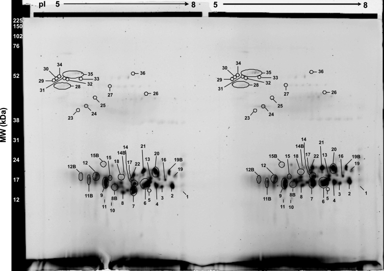
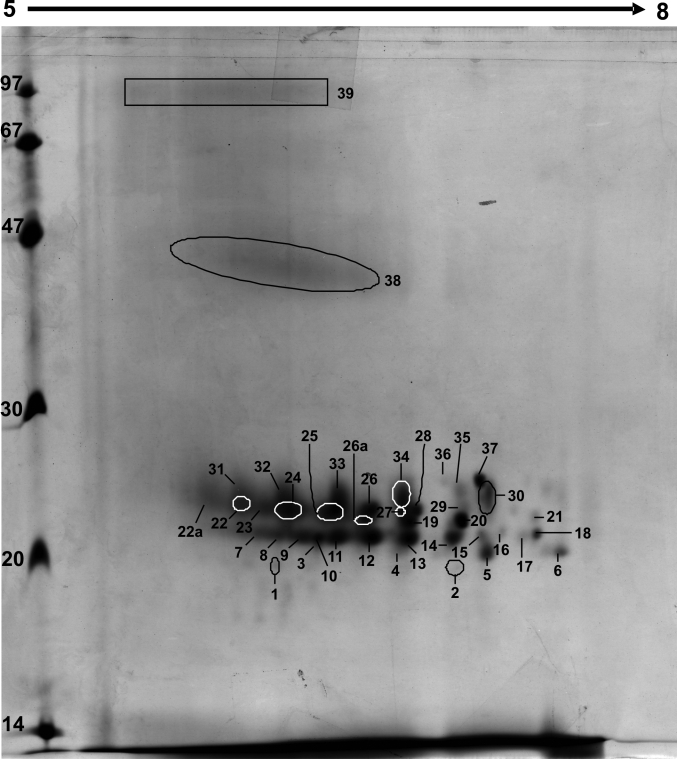
Methods: Laser capture microdissection (LCM) was used to select and capture cells from cortical and nuclear regions of 12 mum, optimum cutting temperature (OCT) compound-embedded frozen lens sections from a 69-year-old human lens. Proteins were extracted and then analyzed by 2-D difference gel electrophoresis (2-D DIGE) with sulfonated indocyanine dye (CyDye) labeling. Crystallin identities and their PTMs were then determined by Matrix-Assisted Laser Desorption/Ionization Time-of-Flight (MALDI-TOF) and Electrospray Ionization Quadripole Linear Ion-Trap Liquid Chromatography (ESI-QTRAP LC-MS/MS) mass spectrometry.
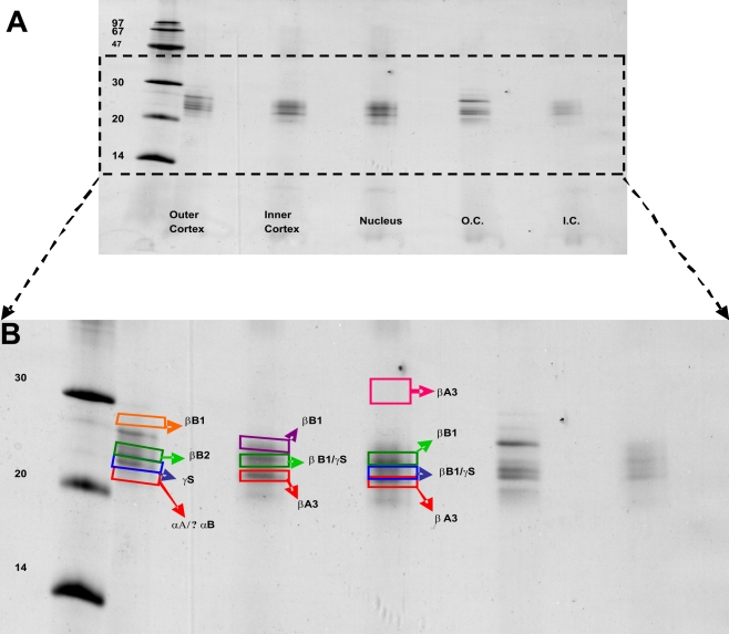
Results: Crystallin fragments (M(r) <20 kDa) were present in both cortical and nuclear regions, while high molecular weight (HMW) aggregates (M(r) > 35 kDa) were mostly localized in the nuclear region. HMW complexes contained a relatively large number of truncated and modified beta-crystallins, compared to alpha- and gamma-crystallins, and two lens-specific intermediate filaments, CP49 (phakinin) and filensin. Modified alpha-crystallins were in low abundance in the nuclear region compared to the cortical region. Several PTMs, including deamidation, oxidation, phosphorylation, ethylation, methylation, acetylation, and carbamylation, were identified in virtually all crystallins and CP49. The data provide the first report of human lens crystallin profiling by a combination of LCM, 2D-DIGE, and mass spectrometric analysis.
Conclusions: The results suggested that as the fiber cells migrate from cortical region to the nuclear region, the crystallin degradation begins in the cortical region and continues in the nuclear region. However, a greater number of the HMW complexes exist mainly in the nuclear region.
Figures
References
-
- Delaye M, Tardieu A. Short-range order of crystallin proteins accounts for eye lens transparency. Nature. 1983;302:415–7. - PubMed
-
- Ponce A, Sorensen C, Takemoto L. Role of short-range protein interactions in lens opacifications. Mol Vis. 2006;12:879–84. - PubMed
-
- Andley UP. Crystallins in the eye: function and pathology. Prog Retin Eye Res. 2007;26:78–98. - PubMed
-
- Bloemendal H, de Jong W, Jaenicke R, Lubsen NH, Slingsby C, Tardieu A. Ageing and vision: structure, stability and function of lens crystallins. Prog Biophys Mol Biol. 2004;86:407–85. - PubMed
-
- Harding JJ. Lens. In: Harding, JJ, editor. Biochemistry of the Eye. London: Chapman & Hall; 1997. p. 94–134.
Publication types
MeSH terms
Substances
Grants and funding
LinkOut - more resources
Full Text Sources
Miscellaneous