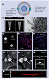
HDL as a contrast agent for medical imaging
- PMID: 20352038
- PMCID: PMC2846093
- DOI: 10.2217/clp.09.38
HDL as a contrast agent for medical imaging
Abstract
Contrast-enhanced MRI of atherosclerosis can provide valuable additional information on a patient's disease state. As a result of the interactions of HDL with atherosclerotic plaque and the flexibility of its reconstitution, it is a versatile candidate for the delivery of contrast-generating materials to this pathogenic lesion. We herein discuss the reports of HDL modified with gadolinium to act as an MRI contrast agent for atherosclerosis. Furthermore, HDL has been modified with fluorophores and nanocrystals, allowing it to act as a contrast agent for fluorescent imaging techniques and for computed tomography. Such modified HDL has been found to be macrophage specific, and, therefore, can provide macrophage density information via noninvasive MRI. As such, modified HDL is currently a valuable contrast agent for probing preclinical atherosclerosis. Future developments may allow the application of this particle to further diseases and pathological or physiological processes in both preclinical models as well as in patients.
Figures


References
-
- Mulder WJM, Cormode DP, Hak S, Lobatto ME, Silvera S, Fayad ZA. Multimodality nanotracers for cardiovascular applications. Nat. Clin. Pract. Cardiovasc. Med. 2008;5:S103–S111. - PubMed
-
- Nakashima Y, Plump AS, Raines EW, Breslow JL, Ross R. ApoE-deficient mice develop lesions of all phases of atherosclerosis through the arterial tree. Arterioscl. Thromb. 1994;14:133–140. - PubMed
-
-
Fayad ZA, Fallon JT, Shinnar M, et al. Noninvasive in vivo high-resolution magnetic resonance imaging of atherosclerotic lesions in genetically engineered mice. Circulation. 1998;98:1541–1547. ▪ MRI visualization of the plaques of atherosclerotic mice.
-
-
- Trogan E, Fayad ZA, Itskovich VV, et al. Serial studies of mouse atherosclerosis by in vivo magnetic resonance imaging detect lesion regression after correction of dyslipidemia. Arterioscler. Thromb. Vasc. Biol. 2004;24:1714–1719. - PubMed
-
- Choudhury RP, Fayad ZA, Aguinaldo JG, et al. Serial, noninvasive, in vivo magnetic resonance microscopy detects the development of atherosclerosis in apolipoprotein E-deficient mice and its progression by arterial wall remodeling. J. Magn. Reson. Imaging. 2003;17:184–189. - PubMed
Grants and funding
LinkOut - more resources
Full Text Sources
Research Materials
