. 2010 Mar 24;5(3):e9872.
doi: 10.1371/journal.pone.0009872.

Mutations in CHMP2B in lower motor neuron predominant amyotrophic lateral sclerosis (ALS)

Affiliations

Mutations in CHMP2B in lower motor neuron predominant amyotrophic lateral sclerosis (ALS)

Laura E Cox et al. PLoS One. .

Abstract

Background: Amyotrophic lateral sclerosis (ALS), a common late-onset neurodegenerative disease, is associated with fronto-temporal dementia (FTD) in 3-10% of patients. A mutation in CHMP2B was recently identified in a Danish pedigree with autosomal dominant FTD. Subsequently, two unrelated patients with familial ALS, one of whom also showed features of FTD, were shown to carry missense mutations in CHMP2B. The initial aim of this study was to determine whether mutations in CHMP2B contribute more broadly to ALS pathogenesis.

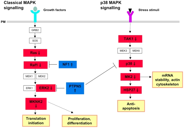
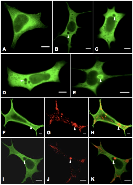
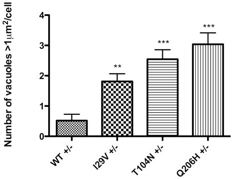
Methodology/principal findings: Sequencing of CHMP2B in 433 ALS cases from the North of England identified 4 cases carrying 3 missense mutations, including one novel mutation, p.Thr104Asn, none of which were present in 500 neurologically normal controls. Analysis of clinical and neuropathological data of these 4 cases showed a phenotype consistent with the lower motor neuron predominant (progressive muscular atrophy (PMA)) variant of ALS. Only one had a recognised family history of ALS and none had clinically apparent dementia. Microarray analysis of motor neurons from CHMP2B cases, compared to controls, showed a distinct gene expression signature with significant differential expression predicting disassembly of cell structure; increased calcium concentration in the ER lumen; decrease in the availability of ATP; down-regulation of the classical and p38 MAPK signalling pathways, reduction in autophagy initiation and a global repression of translation. Transfection of mutant CHMP2B into HEK-293 and COS-7 cells resulted in the formation of large cytoplasmic vacuoles, aberrant lysosomal localisation demonstrated by CD63 staining and impairment of autophagy indicated by increased levels of LC3-II protein. These changes were absent in control cells transfected with wild-type CHMP2B.

Conclusions/significance: We conclude that in a population drawn from North of England pathogenic CHMP2B mutations are found in approximately 1% of cases of ALS and 10% of those with lower motor neuron predominant ALS. We provide a body of evidence indicating the likely pathogenicity of the reported gene alterations. However, absolute confirmation of pathogenicity requires further evidence, including documentation of familial transmission in ALS pedigrees which might be most fruitfully explored in cases with a LMN predominant phenotype.


Conflict of interest statement

Competing Interests: The authors have declared that no competing interests exist.

Figures

Figure 1
Figure 1. Chromatograms showing nucleotide changes in CHMP2B.
On the left side of each image is the normal wild-type sequence, whilst the right side shows the nucleotide change for each of the changes identified in CHMP2B.
Figure 2
Figure 2. Photomicrography of pathological changes associated with CHMP2B mutations.
In all cases there was no evidence of myelin pallor affecting the corticospinal tracts (a). Case 1 showed some minor upregulation of CD68 immunoreactivity in the spinal lateral corticospinal tracts (b) compared with the dorsal columns (c). Sequestosome 1/p62 staining showed compact intraneuronal inclusions in spinal motor neurons and occasional glial inclusions (d). The glial inclusions show coiled body morphology immunoreactive for both TDP43 (e) and p62 (f). All cases showed a predominance of compact intraneuronal inclusions in motor neurons (g–i). (a: Luxol fast blue; b,c: CD68; d,f,h,i: p62; e,g: TDP43. Microscopy at ×2 obj. (a); ×10 obj. (b,c); ×40 obj. (d–i)).
Figure 3
Figure 3. Summary of key gene expression changes to ER and Golgi function, vesicular transport, mTOR signalling and autophagy.
The downregulation of multiple transcripts encoding ribosomal proteins, translation initiation factors (eIFs) and the ribosomal subunit, RPS6 suggests a global repression of translation within the cell (1). SEC23 and SEC24 coat vesicles into which immature proteins are packaged and are upregulated. Vesicles are transported along microtubules (MTs) from the ER-Golgi intermediate compartment; however, downregulation of the main components of microtubules, TUBA and TUBB, and the MT-stabilising proteins, MAP1S and MAP4, indicates MT disassembly and therefore disruption to vesicular transport (2). This is enhanced by upregulation of STMN1, a MT-destabilising protein, whose over-expression has also been demonstrated to result in Golgi fragmentation. Downregulation of transcripts maintaining Golgi structure (GLG1, GORASP1, COG2 and COG7) support the hypothesis of Golgi fragmentation (3). COPI is used to coat empty vesicles exiting the Golgi for recycling back to the ER, however, two main constituents, COPE and COPZ1, are downregulated, as is USE1, which is required for vesicle fusion with the ER. These findings predict an eventual deficit of material available to ER for the packaging of newly synthesised proteins (4). There is dysregulation of multiple SNARE transcripts, which are required for vesicle fusion. BOS1 and SEC22 are upregulated, which may be the Golgi's response to the reduction in vesicles being transported along destabilised microtubules. Multiple SNAREs and adaptor proteins (VTI1, STX4, AP1B1, AP2A2, AP2S1, AP3D1 and AP4B1), which are required for fusion between vesicles carrying mature proteins and the cell surface, endosomes and lysosomes, are downregulated, predicting impairment in the delivery of proteins throughout the cell (5). Finally, inhibition of autophagy by the mTORC1 complex and downregulation of ATG1, which forms the phagophore assembly site (PAS) with ATG17 and ATG13 to initiate autophagy, indicates a decrease in the clearance of cellular debris which may result in cytosolic accumulations and contribute to motor neuron injury (6).
Figure 4
Figure 4. Defects in MAPK signalling as a result of mutations in CHMP2B.
There are two main pathways through which MAP kinases signal: the classical MAPK pathway and the p38 MAP kinase pathway. In the classical signalling pathway, ligand binding results in receptor activation, which in turns leads to the activation of GRB2 and SOS. SOS catalyses the substitution of GDP for GTP on RAS (⇓), which then activates RAF1 (⇓). RAF1 subsequently phosphorylates MEK1/2, which in turn activate ERK1 and ERK2 (⇓). The ERK proteins activate MKNK2 (⇓), which is directly responsible for activation of proteins required for translation initiation. Downregulation of multiple core signalling components in combination with upregulation of the inhibitors, NF1 and PTPN5 predicts inability of the cell's basal response to growth factors. The p38 MAPK pathway is a signalling cascade that is distinct, but not exclusive from the classical MAPK signalling pathway. As with the classical MAPK pathway, multiple elements of the pathway are downregulated: TAK1(⇓), p38 (⇓), MK2 (⇓) and HSP27 (⇓) which may result in a decrease in mRNA stability and the cell's anti-apoptotic response. (PM = plasma membrane)
Figure 5
Figure 5. Defects in calcium signalling as a result of CHMP2B mutations.
Downregulation of the α-subunits, Gq (⇓) and Gq11 (⇓) indicates a decrease in PLCβ activation, despite upregulation of the cholinergic receptor CHRM3 (⇑). A decrease in PLCβ activity would reduce the amount of phosphatidylinositol 4,5-bisphosphate (PIP2) hydrolysed to inositol 1,4,5-triphosphate (IP3) and diacylglycerol (DAG). Combining the decrease in IP3, with downregulation of its receptor, IP3R (⇓), would reduce the opening of a Ca2+-release channel on the ER membrane, which allows Ca2+ to exit the ER lumen into the cytosol. SERCA, which pumps Ca2+ out of the cytoplasm into the ER lumen is upregulated, so these findings predict an increased Ca2+ concentration in the ER lumen. Upregulation of NCX1(⇑) and VDAC3(⇑), and downregulation of ANT3(⇓), predict that mitochondria are increasing the amount of calcium pumped out of the matrix, but decreasing the amount of ATP leaving the organelle. Amplifying the aberrant intracellular calcium levels, in addition to reducing the amount of energy available for cellular processes. (PM = plasma membrane; OMM = outer mitochondrial matrix; IMM = inner mitochondrial matrix).
Figure 6
Figure 6. Overexpression of mutant CHMP2B produces an aberrant phenotype in HEK-293 cells.
Cells were transfected with vectors encoding recombinant protein c-Myc-CHMP2B with either the wild-type or I29V, T104N or Q206H mutant sequence, and stained with FITC-conjugated antibody to c-Myc. Transfection with wild-type CHMP2B (A) results in generalised cytoplasmic expression, whereas the mutant isoforms I29V (B), T104N (C) and Q206H (D) resulted in cytoplasmic vacuoles of varying size (indicated by arrowheads). Another striking observation was the presence within cells expression mutant CHMP2B of circular CHMP2B accumulations in the cytoplasm, termed halos (E). Cells were doubly stained with antibodies to c-Myc (F & I), as well as CD63 (G & J), and merged to show co-localisation (H & K). CD63 co-localises with the small vacuoles found in cells transfected with WT CHMP2B (F–H). However, CD63 staining does not co-localise with large vacuoles in mutant expressing cells (cells transfected with T104N shown), but are found on the vacuole edge (I–K). Images were taken on a Zeiss LSM 510 confocal microscope, ×63 obj. Bar, 10µm.
Figure 7
Figure 7. Mutant CHMP2B causes large cytoplasmic vacuoles.
Cells expressing 3 different mutant CHMP2B had significantly more vacuoles with an area greater than 1µm2 in than cells expressing wild-type protein (One-way ANOVA p<0.0001, +/−: cells grown in DMEM with FCS, without antibiotic).
Figure 8
Figure 8. LC3-II levels are increased in cells expressing mutant CHMP2B.
A representative image of western blotting to LC3 and actin (A), showing increased levels of LC3-II in COS-7 cells expressing mutant CHMP2B protein (lanes 2–4) compared to cells expressing wild-type protein (lane 1). LC3-II levels relative to an actin loading control were measured using densitometry (B), and this showed increased levels of LC3-II in mutant expressing cells compared to WT (mean ±S.E.M., n = 3) (Mann Whitney p = 0.0242). Average fold changes in LC3-II levels normalised to WT are shown for each of the three mutants (C).

References

    1. Shaw PJ. Molecular and cellular pathways of neurodegeneration in motor neurone disease. J Neurol Neurosurg Psychiatry. 2005;76:1046–1057. - PMC - PubMed
    1. Siddique N, Siddique T. Genetics of amyotrophic lateral sclerosis. Phys Med Rehabil Clin N Am. 2008;19:429–439, vii. - PMC - PubMed
    1. Parkinson N, Ince PG, Smith MO, Highley R, Skibinski G, et al. ALS phenotypes with mutations in CHMP2B (charged multivesicular body protein 2B). Neurology. 2006;67:1074–1077. - PubMed
    1. Skibinski G, Parkinson NJ, Brown JM, Chakrabarti L, Lloyd SL, et al. Mutations in the endosomal ESCRTIII-complex subunit CHMP2B in frontotemporal dementia. Nat Genet. 2005;37:806–808. - PubMed
    1. Babst M, Katzmann DJ, Estepa-Sabal EJ, Meerloo T, Emr SD. Escrt-III: an endosome-associated heterooligomeric protein complex required for mvb sorting. Dev Cell. 2002;3:271–282. - PubMed

Publication types

MeSH terms

Substances