. 2010 Mar 24;5(3):e9850.
doi: 10.1371/journal.pone.0009850.

The MinCDJ system in Bacillus subtilis prevents minicell formation by promoting divisome disassembly

Affiliations

The MinCDJ system in Bacillus subtilis prevents minicell formation by promoting divisome disassembly

Suey van Baarle et al. PLoS One. .

Abstract

Background: Cell division in Bacillus subtilis takes place precisely at midcell, through the action of Noc, which prevents division from occurring over the nucleoids, and the Min system, which prevents cell division from taking place at the poles. Originally it was thought that the Min system acts directly on FtsZ, preventing the formation of a Z-ring and, therefore, the formation of a complete cytokinetic ring at the poles. Recently, a new component of the B. subtilis Min system was identified, MinJ, which acts as a bridge between DivIVA and MinCD.

Methodology/principal findings: We used fluorescence microscopy and molecular genetics to examine the molecular role of MinJ. We found that in the absence of a functional Min system, FtsA, FtsL and PBP-2B remain associated with completed division sites. Evidence is provided that MinCDJ are responsible for the failure of these proteins to localize properly, indicating that MinCDJ can act on membrane integral components of the divisome.

Conclusions/significance: Taken together, we postulate that the main function of the Min system is to prevent minicell formation adjacent to recently completed division sites by promoting the disassembly of the cytokinetic ring, thereby ensuring that cell division occurs only once per cell cycle. Thus, the role of the Min system in rod-shaped bacteria seems not to be restricted to an inhibitory function on FtsZ polymerization, but can act on different levels of the divisome.


Conflict of interest statement

Competing Interests: The authors declare that no competing interests exist.

Figures

Figure 1
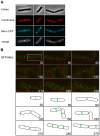
Figure 1. Dynamic localization of MinJ.
A. Localization of MinJ-CFP, expressed from its native locus (strain SB003). From top to bottom the images show the phase contrast, membrane stain (FM4-64), MinJ-CFP, and merged image of membrane and MinJ-CFP. The scale bar is 5 µm. Three different localization patterns are shown: on the left panel, MinJ-CFP localizes at young poles/midcell (44.6%), in the middle panel, MinJ-CFP localizes to both poles (37.6%), and in the right panel, MinJ-CFP localizes to both midcell and poles (17.8%). In total 250 cells were counted. B. Time lapse microscopy of GFP-MinJ (strain MB001) showing the dynamic localization of MinJ. Numbers indicate minutes. Top shows a merged image of phase contrast and GFP-MinJ microscopy image, the bottom part shows a cartoon of localization of GFP-MinJ of one particular cell (highlighted in white box in microscopy image). The image shows that the localization of GFP-MinJ depends on the state of the cell cycle. When cells are not dividing, GFP-MinJ is localized to the poles. As cells prepare to divide, GFP-MinJ moves from the poles to midcell. After division is completed, GFP-MinJ moves back to the poles. The complete movie can be seen in the supplemental material (Movie S2).
Figure 2
Figure 2. FtsA-YFP remains associated with the cell poles in the absence of a functional Min system.
A. From top to bottom, localization of FtsA-YFP in wild type cells (SB067), ΔminCD (SB060), ΔminJ (SB066), and ΔminCDJ (SB061). Left to right: phase contrast image, membrane stain (FM4-64), FtsA-YFP, and a merged image of the membrane stain and FtsA-YFP. Scale bar is 5 µm. Arrows indicate FtsA rings resembling spirals, which are found at cell poles/late septa. B. Percentage of cell poles containing FtsA-YFP in wild type cells (SB067), ΔminCD (SB060), ΔminJ (SB066), and ΔminCDJ (SB061) (n = 200).
Figure 3
Figure 3. Time-lapse microscopy of FtsA-YFP.
On the left, FtsA-YFP in wild type (SB067), center: in ΔminJ (SB066) and right in ΔminCD (SB060). Numbers on the left indicate minutes and numbers on the FtsA-YFP images show the generation of the rings. In wild type, ring 1 rapidly disappears and new rings are formed at midcell of the two progeny cells (rings 2) which also disappear after division is complete, while new rings again appear at midcell. In the absence of MinJ, the FtsA-YFP ring in this strain (ring 1) is not disassembled and instead begins forming double rings (rings 2). The same was observed for ΔminCD. The merge image is an overlay of the phase contrast image with the corresponding FtsA-YFP signal.
Figure 4
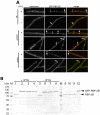
Figure 4. GFP-PBP-2B remains at the cell pole in Δmin cells.
A. Shown are strains SB088 (GFP-PBP-2B), SB092 (GFP-PBP-2B ΔminD), and SB090 (GFP-PBP-2B ΔminJ) grown with (+) and without (−) 1 mM IPTG (pre-cultures were grown with 1 mM IPTG). From left to right is shown the membrane stain, GFP-PBP-2B and a merged image. Scale bar is 5 µm. B. Western blot of different ftsZ/min mutants with α-PBP-2B. The loading pattern of the different lanes is as follows: 1/5: FtsZ+ (strain 1801), 2/6: FtsZ+ GFP-PBP-2B+ (strain SB088), 3/7: FtsZ+ ΔminD GFP-PBP-2B+ (strain SB090), 4/8: FtsZ+ ΔminJ GFP-PBP-2B+ (strain SB092), 9: wild type (strain 168), 10: ΔminJ (strain RD021), 11: ΔminDJ (strain SB075), 12: GFP-PBP-2B (strain 3122). Lanes with molecular mass standard are labelled with M. Induction of GFP-PBP-2B was done with 0.5% xylose and FtsZ expression was induced with 1 mM IPTG (FtsZ+) or depleted (FtsZ). Note that a full-length GFP-PBP-2B band is at 106.2 kDA and the native PBP-2B band is seen at 79.1 kDa.
Figure 5
Figure 5. Cells without a functional Min system form multiple minicells.
Shown are examples for ΔminCD (3309), ΔminCJ (SB074), ΔminDJ (SB075), and ΔminCDJ (MB012), with phase images and membrane stains taken for a few exemplary cells. Note the formation of 2–4 minicells in a row, indicating that a divisome that fails to disassemble often initiates a new round of division, resulting in multiple minicell formation. Scale bar is 5 µm.
Figure 6
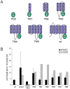
Figure 6. MinJ is able to modulate MinCD activity.
A. A series of MinJ truncations were created to test which domains are important for function. Note that all truncation were expressed as C-terminal GFP fusions. Localization of the fusion proteins can be found in supplemental material Fig. S5. These truncations include the soluble PDZ domain, TM1, containing the PDZ domain and the last transmembrane helix; TM2, with the PDZ domain and the last two transmembrane helices, and so forth. B. To test functionality, they were expressed in ΔminJ cells and the cell length and amount of minicells were measured. From left to right, strains tested were wild type (168), ΔminJ (RD021), ΔminJ MinJ+ (MB004), PDZ (SB010), TM1 (SB004), TM2 (SB005), TM3 (SB006), TM4 (SB007) and TM5 (SB008). Grey bars indicate the percentage of minicells produced, and the black bars indicate the average cell length. Expression of TM1 and TM2 led to an identical length as wild type, although they produced even more minicells than the MinJ knockout.
Figure 7
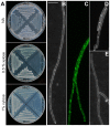
Figure 7. MinD-GFP overexpression increases the cell length.
A. Nutrient agar plates containing, from top to bottom, 0, 0.5%, and 1% xylose inoculated with strains MinD+ (SB076), MinD+ ΔminJ (SB078), MinC+ (SB080), MinC+ ΔminJ (SB082). Cells overexpressing MinD in ΔminJ have a growth defect and grow with difficulty on nutrient agar plates supplemented with 0.5 and 1% xylose. B. MinD overexpression in ΔminJ (SB078) results in extremely long filamentous cells. C. MinD-GFP localizes in foci all over the cell when overexpressed (with 1% xylose) in ΔminJ background (SB052). D. MinD overexpression in wild type (SB076) leads to weak filamentation, although many cells are of normal length. E. MinC overexpression in ΔminJ (SB082) does not lead to any increased filamentation (see also Table 1). Scale bar is 5 µm.
Figure 8
Figure 8. Effects of MinD overexpression on MinC and FtsA.
A. Left, MinC-GFP localization in wild type (EBS499), center: in MinD+ (SB086) induced with 1% xylose, and right, in ΔminJ (SB086). top to bottom, phase contrast, membrane stain, MinC-GFP, and a merged image of the membrane stain and MinC-GFP. MinD overexpression leads to a localization pattern of MinC-GFP with multiple rings forming throughout the cell, with double rings frequently being observed. In the absence of MinJ and overexpression of MinD, MinC-GFP becomes completely dispersed and forms foci throughout the cell. B. Left: FtsA-YFP localization in MinD+ (SB084) and right: in MinD+ ΔminJ (SB085). In both strains expression of MinD was induced with 1% xylose. From top to bottom, the figure shows an image of the membrane stain (FM4-64), FtsA-YFP localization, and a merged image of FtsA-YFP and the membrane stain. FtsA-YFP expressed in cells overexpressing MinD still localizes in wild type and ΔminJ cells indicating that the filamentous cell phenotype must occur downstream of FtsA recruitment to the Z-ring.

Similar articles

Cited by

References

    1. Harry E, Monahan L, Thompson L. Bacterial cell division: the mechanism and its precison. Int Rev Cytol. 2006;253:27–94. - PubMed
    1. Goehring NW, Beckwith J. Diverse paths to midcell: assembly of the bacterial cell division machinery. Curr Biol. 2005;15:R514–526. - PubMed
    1. Errington J, Daniel RA, Scheffers DJ. Cytokinesis in bacteria. Microbiol Mol Biol Rev. 2003;67:52–65, table of contents. - PMC - PubMed
    1. Adams DW, Errington J. Bacterial cell division: assembly, maintenance and disassembly of the Z ring. Nat Rev Microbiol. 2009;7:642–653. - PubMed
    1. Bi EF, Lutkenhaus J. FtsZ ring structure associated with division in Escherichia coli. Nature. 1991;354:161–164. - PubMed

Publication types

MeSH terms