. 2010 Mar 23;5(3):e9832.
doi: 10.1371/journal.pone.0009832.

Transient tumor-fibroblast interactions increase tumor cell malignancy by a TGF-Beta mediated mechanism in a mouse xenograft model of breast cancer

Affiliations

Transient tumor-fibroblast interactions increase tumor cell malignancy by a TGF-Beta mediated mechanism in a mouse xenograft model of breast cancer

Christina H Stuelten et al. PLoS One. .

Abstract

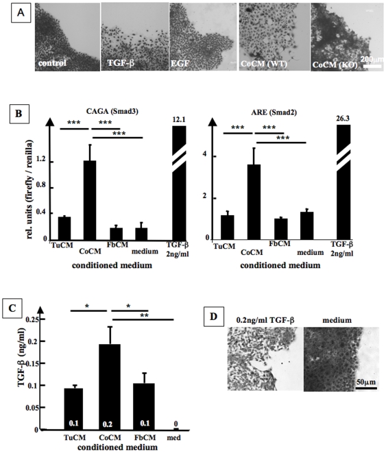
Carcinoma are complex societies of mutually interacting cells in which there is a progressive failure of normal homeostatic mechanisms, causing the parenchymal component to expand inappropriately and ultimately to disseminate to distant sites. When a cancer cell metastasizes, it first will be exposed to cancer associated fibroblasts in the immediate tumor microenvironment and then to normal fibroblasts as it traverses the underlying connective tissue towards the bloodstream. The interaction of tumor cells with stromal fibroblasts influences tumor biology by mechanisms that are not yet fully understood. Here, we report a role for normal stroma fibroblasts in the progression of invasive tumors to metastatic tumors. Using a coculture system of human metastatic breast cancer cells (MCF10CA1a) and normal murine dermal fibroblasts, we found that medium conditioned by cocultures of the two cell types (CoCM) increased migration and scattering of MCF10CA1a cells in vitro, whereas medium conditioned by homotypic cultures had little effect. Transient treatment of MCF10CA1a cells with CoCM in vitro accelerated tumor growth at orthotopic sites in vivo, and resulted in an expanded pattern of metastatic engraftment. The effects of CoCM on MCF10CA1a cells were dependent on small amounts of active TGF-beta1 secreted by fibroblasts under the influence of the tumor cells, and required intact ALK5-, p38-, and JNK signaling in the tumor cells. In conclusion, these results demonstrate that transient interactions between tumor cells and normal fibroblasts can modify the acellular component of the local microenvironment such that it induces long-lasting increases in tumorigenicity and alters the metastatic pattern of the cancer cells in vivo. TGF-beta appears to be a key player in this process, providing further rationale for the development of anti-cancer therapeutics that target the TGF-beta pathway.


Conflict of interest statement

Competing Interests: The authors have declared that no competing interests exist.

Figures

Figure 1
Figure 1. Cocultures of breast cancer cells are well organized and increase motility of tumor cells.
A. Upper panel. Human breast cancer cells (CA1a) and mouse dermal fibroblasts (DF) form a well organized 2D coculture where fibroblast streaks surround tumor cell islets. Middle panel. Use of fibroblasts and CA1a cells that report Smad3 dependent TGF-β signaling by expression of RFP and GFP, respectively, demonstrates that TGF-β signaling in both cell types is increased in cocultures as compared to the homotypic cultures. Bottom Panel. Outline of the tumor cell and fibroblast compartments of the images shown in the middle panel. B. Medium conditioned by cocultures (CoCM) as compared to medium from homotypic fibroblast (FbCM) and tumor cell (TuCM) cultures increases cell migration and cell scattering in scratch assays (24 h) and dot assays (4 d), and induces cytoplasmic localization of E-cadherin as visualized by confocal microscopy (60 min). C. Influence of CoCM on closure of in vitro wounds (“scratch assay”). Cells were plated to confluence, mitosis inhibited by preincubation with mitomycin, and cells then stimulated with conditioned media. CoCM causes significantly faster closure of scratches as compared to all other conditioned media (scratch width after 24 h, n = 8 samples/group, ANOVA/Bonferroni; p<0.0001). D. Quantification of E-cadherin related immunofluorescence in CA1a cells treated with CM (Fig. 1B). Membranes and cytoplasm of cells were gated separately and average signal intensity was determined using ImageJ. Ratios of membrane and cytoplasmic signal were analyzed for statistical significance using GraphPad Prism (Kruskal-Wallis test/Dunn's Multiple Comparison Test, p = 0.0052).
Figure 2
Figure 2. TGF-β signaling induces scattering of CA1a cells.
A. Exogenous TGF-β and fibroblast-released TGF-β induces cell scattering in dot assays. Cells were plated into dot assays, incubated over night, and then stimulated with TGF-β (5 ng/ml), EGF (100 ng/ml), or vehicle (Control) for 4 d. Use of CoCM that was derived from cocultures of CA1a cells and TGFβ1 knockout fibroblasts (CoCM(KO)) did not stimulate cell scattering while conditioned medium from cocultures employing TGF-β1 wildtype fibroblasts (CoCM(WT)) did induce cell scattering. B. CoCM as compared to TuCM and FbCM induces higher levels of CAGA (Smad3) and ARE (Smad2) mediated luciferase activity in CA1a cells (n = 3, ANOVA/Dunnett's Multiple Comparison, p = 0.0002 (ARE) and p<0.0001 (CAGA)). C. CoCM contains higher levels of TGF-β than TuCM and FbCM. CCL-64 cells were stimulated with CM overnight and PAI-driven luciferase activity was measured. D. Scattering of CA1a cells is induced by addition of 0.2 ng/ml TGF-β to medium. Cells were plated into dot assays, incubated overnight, and then stimulated with TGF-β (0.2 ng/ml) or vehicle for 4 d.
Figure 3
Figure 3. CoCM activates Smad2/3-signaling and the MAPK signaling cascades p38 and JNK in CA1a cells suggesting activation of canonical and noncanonical TGF-β signaling by CoCM derived TGF-β.
A. TGF-β activates canonical signaling via Smad2/3 and non-canonical signaling via TAK-1 relayed JNK- and p38 signaling cascades. CoCM stimulated both, phosphorylation of TAK1, and phosphorylation of Smad2/Smad3. Cells were incubated with CoCM for 30 min (pTAK1) or 60 min (pSmad2, pSmad3). B. CoCM as compared to TuCM and FbCM induces nuclear localization of Smad2/3 within 60 min, indicating activation of canonical Smad signaling upon stimulation with CoCM. C. Quantification of Smad2/3 mediated immunofluorescence in CM treated CA1a cells (Fig. 2B). Nuclei and cytoplasm of cells were gated separately and average signal intensity was determined using ImageJ. Ratios of nuclear and cytoplasmic signal intensities were analyzed for statistical significance using GraphPad Prism (ANOVA/Dunnett's Multiple Comparison Test, p<0.0001, n = number of cells analyzed).
Figure 4
Figure 4. CoCM activates the MAPK signaling cascades p38 and JNK signaling in CA1a.
A. Stimulation of CA1a cells with CoCM as compared to FbCM and TuCM for 60 min increases levels of phospho-p38 (pp38), pATF-2, pMAPKAPK-2, and pJNK as visualized by fluorescence microscopy, indicating that CoCM activates these non-canonical TGF-β signaling cascades. For quantification of pp38-, pATF-2-, pMAPKAPK2- and pJNK mediated immunofluorescence cells were gated and average signal intensity was determined using ImageJ. Ratios of nuclear and cytoplasmic signal intensities were analyzed for statistical significance using GraphPad Prism (Kruskal-Wallis test/Dunn's Multiple Comparison Test, p<0.0001 [pp38], p<0.0001 [pATF-2], p<0.0001 [pMAPKAPK2], p<0.00001 [pJNK]). 30 to 40 cells were analyzed per group. B. Inhibitors were added to cultures 30 min prior to stimulating CA1a cells with CoCM for 60 min. E-cadherin was visualized by immunocytochemistry and confocal microscopy. Nuclei were stained with DAPI. For quantification of E-cadherin mediated the cell membrane and the cytoplasm were gated for each cell and average signal intensities were determined using ImageJ. Ratios of nuclear and cytoplasmic signal intensities were analyzed for statistical significance using GraphPad Prism (ANOVA/Dunnetts's Multiple Comparison Test, p<0.0001. n  =  number of cells analyzed in each group.
Figure 5
Figure 5. CoCM increases tumorigenicity and expands the metastatic pattern of CA1a cells.
A. Experimental design of orthotopic implantation- and tail vein injection models. CA1a cells were pre-treated ex vitro with CM for 4 days, trypsinized, washed, and resuspended in DPBS. Orthotopic Implantation Model. 4×10⁴ cells in 50 µl DPBS were bilaterally injected into the axillary mammary fat pads of female SCID mice (15 animals or 30 tumor inoculation sites/group). Tumor size was assessed thrice weekly using calipers. Estimated tumor volumes were calculated by the formula (S×S×L)×0.52, where S and L are the short and long dimensions, respectively . Tail Vein Injection Model. 2×10⁵ cells in 100 µl DPBS were injected into the tail vein of female NOD SCID mice, and animals were monitored twice weekly for signs of metastasis. B. Ex vivo pre-treatment of CA1a cells with CoCM accelerates growth of orthotopic xenograft tumors in vivo. The axillary mammary fat pads of female SCID mice were bilaterally inoculated with CA1a cells that were pre-treated with CM (15 animals or 30 tumor inoculation sites/group). Tumor volumes at day 23 were analyzed using GraphPad Prism 5.0. Kruskal Wallis test/Dunn's multiple comparison; p<0.001; CoCM vs TuCM: ***, CoCM vs FbCM: **, CoCM vs medium: ***, TuCM vs FbCM: N.S., TuCM vs medium: N.S., FbCM vs medium: N.S.. Representative of 2 independent experiments. C. Ex vivo pre-treatment of CA1a cells with CoCM results in extrapulmonary metastases in tail vein injection assays. CA1a cells were incubated with conditioned media for 4 days and injected into the tail vein of female NOD SCID mice. While all animals developed lung metastases, only animals injected with CoCM treated CA1a cells developed extrapulmonary tumors. Extrapulmonary tumor growth was suppressed when TGF-β signaling was inhibited by the ALK 5 inhibitor SB431542 during exposure of CA1a cells to CoCM. Animals receiving CoCM pre-treated cells had significantly higher occurrence of extrapulmonary tumors than all other groups (Chi Square Test, tail vein injection #1: p = 0.0002; tail vein injection #2: p = 0.0085). D. Functional TGF-β signaling of tumor cells is required for CoCM induced formation of extrapulmonary metastases. Blocking of ALK5 mediated TGF-β signaling by the ALK5-inhibitor SB435142 during in vitro exposure of CA1a cells to CoCM inhibited metastases and significantly increased survival time of animals in vivo (median survival: CoCM: 62 days (n = 13 animals), SB431542: 96 days (n = 14 animals), Kaplan Maier Analysis, p<0.0001).
Figure 6
Figure 6. CoCM expands the metastatic pattern of CA1a cells and induces sustained TGF-β signaling via pSmad2.
A. Extrapulmonary tumors formed by CoCM pre-treated CA1a cells after tail vein injection were highly invasive. Extrapulmonary tumors were observed in the subcutaneously, often in the proximity to mammary fat pads (arrows indicate a normal mammary duct, hematoxylin-eosin staining). B. Xenograft tumors and extrapulmonary metastatic tumors derived from CoCM pre-treated cells have higher levels of nuclear pSmad2 than xenograft tumors derived from medium pre-treated CA1a cells as shown by immunostaining. Levels of pSmad2 in xenograft and metastatic tumors were higher in CA1a cells invading the surrounding tissue (arrows) as compared to tumor cells in the center of tumors. The negative control was obtained by omitting the primary antibody. C. Nuclear pSmad2 is increased in tumor cells at the tumor stroma border (arrows) as compared to cells in the center of tumors that spontaneously developed in a C3(1)TAg model of breast cancer and in an orthotopic syngeneic model of breast cancer (4T1 cells/BALB/c mice).
Figure 7
Figure 7. Tumor-stroma interactions increase malignancy of tumor cells by bidirectional effects of TGF-β.
Direct interactions of tumor cells and fibroblasts as they occur when parenchymal tumor cells invade the underlying stroma increase secretion of TGF-β by tumor cells as well as by fibroblasts. TGF-β has bidirectional effects on both cells types. Fibroblast derived TGF-β can directly stimulate tumor cell migration and malignancy. Tumor cell derived TGF-β induces fibroblasts to secrete MMP-9 . Increased MMP-9 levels then might further increase levels of active TGF-β, and also facilitate matrix degradation and migration of tumor cells, further increasing the malignant potential of tumor cells.

Similar articles

Cited by

References

    1. Beacham DA, Cukierman E. Stromagenesis: the changing face of fibroblastic microenvironments during tumor progression. SeminCancer Biol. 2005;15:329–341. - PubMed
    1. Proia DA, Kuperwasser C. Stroma: tumor agonist or antagonist. Cell Cycle. 2005;4:1022–1025. - PubMed
    1. Olumi AF, Grossfeld GD, Hayward SW, Carroll PR, Tlsty TD, et al. Carcinoma-associated fibroblasts direct tumor progression of initiated human prostatic epithelium. Cancer Res. 1999;59:5002–5011. - PMC - PubMed
    1. Liotta LA, Kohn EC. The microenvironment of the tumour-host interface. Nature. 2001;411:375–379. - PubMed
    1. Baglole CJ, Ray DM, Bernstein SH, Feldon SE, Smith TJ, et al. More than structural cells, fibroblasts create and orchestrate the tumor microenvironment. ImmunolInvest. 2006;35:297–325. - PubMed

Publication types

Substances