C/EBPalpha and C/EBPbeta are required for Sebocyte differentiation and stratified squamous differentiation in adult mouse skin
- PMID: 20352127
- PMCID: PMC2843749
- DOI: 10.1371/journal.pone.0009837
C/EBPalpha and C/EBPbeta are required for Sebocyte differentiation and stratified squamous differentiation in adult mouse skin
Abstract
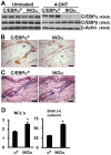
C/EBPalpha and C/EBPbeta are bZIP transcription factors that are highly expressed in the interfollicular epidermis and sebaceous glands of skin and yet germ line deletion of either family member alone has only mild or no effect on keratinocyte biology and their role in sebocyte biology has never been examined. To address possible functional redundancies and reveal functional roles of C/EBPalpha and C/EBPbeta in postnatal skin, mouse models were developed in which either family member could be acutely ablated alone or together in the epidermis and sebaceous glands of adult mice. Acute removal of either C/EBPalpha or C/EBPbeta alone in adult mouse skin revealed modest to no discernable changes in epidermis or sebaceous glands. In contrast, co-ablation of C/EBPalpha and C/EBPbeta in postnatal epidermis resulted in disruption of stratified squamous differentiation characterized by hyperproliferation of basal and suprabasal keratinocytes and a defective basal to spinous keratinocyte transition involving an expanded basal compartment and a diminished and delayed spinous compartment. Acute co-ablation of C/EBPalpha and C/EBPbeta in sebaceous glands resulted in severe morphological defects, and sebocyte differentiation was blocked as determined by lack of sebum production and reduced expression of stearoyl-CoA desaturase (SCD3) and melanocortin 5 receptor (MC5R), two markers of terminal sebocyte differentiation. Specialized sebocytes of Meibomian glands and preputial glands were also affected. Our results indicate that in adult mouse skin, C/EBPalpha and C/EBPbeta are critically involved in regulating sebocyte differentiation and epidermal homeostasis involving the basal to spinous keratinocyte transition and basal cell cycle withdrawal.
Conflict of interest statement
Figures


Similar articles
-
Expression of CCAAT/enhancer binding proteins (C/EBP) is associated with squamous differentiation in epidermis and isolated primary keratinocytes and is altered in skin neoplasms.J Invest Dermatol. 1998 Jun;110(6):939-45. doi: 10.1046/j.1523-1747.1998.00199.x. J Invest Dermatol. 1998. PMID: 9620302
-
C/EBPalpha and beta couple interfollicular keratinocyte proliferation arrest to commitment and terminal differentiation.Nat Cell Biol. 2009 Oct;11(10):1181-90. doi: 10.1038/ncb1960. Epub 2009 Sep 13. Nat Cell Biol. 2009. PMID: 19749746
-
Genetic ablation of CCAAT/enhancer binding protein alpha in epidermis reveals its role in suppression of epithelial tumorigenesis.Cancer Res. 2007 Jul 15;67(14):6768-76. doi: 10.1158/0008-5472.CAN-07-0139. Cancer Res. 2007. PMID: 17638888 Free PMC article.
-
Peroxisome proliferator-activated receptors and skin development.Horm Res. 2000;54(5-6):269-74. doi: 10.1159/000053270. Horm Res. 2000. PMID: 11595816 Review.
-
Lipid droplets and associated proteins in sebocytes.Exp Cell Res. 2016 Jan 15;340(2):205-8. doi: 10.1016/j.yexcr.2015.11.008. Epub 2015 Nov 11. Exp Cell Res. 2016. PMID: 26571075 Review.
Cited by
-
Membrane-bound and exosomal metastasis-associated C4.4A promotes migration by associating with the α(6)β(4) integrin and MT1-MMP.Neoplasia. 2012 Feb;14(2):95-107. doi: 10.1593/neo.111450. Neoplasia. 2012. PMID: 22431918 Free PMC article.
-
C/EBPβ suppresses keratinocyte autonomous type 1 IFN response and p53 to increase cell survival and susceptibility to UVB-induced skin cancer.Carcinogenesis. 2019 Sep 18;40(9):1099-1109. doi: 10.1093/carcin/bgz012. Carcinogenesis. 2019. PMID: 30698678 Free PMC article.
-
The C/EBPβ LIP isoform rescues loss of C/EBPβ function in the mouse.Sci Rep. 2018 May 30;8(1):8417. doi: 10.1038/s41598-018-26579-y. Sci Rep. 2018. PMID: 29849099 Free PMC article.
-
C/EBPβ deficiency enhances the keratinocyte innate immune response to direct activators of cytosolic pattern recognition receptors.Innate Immun. 2023 Jan;29(1-2):14-24. doi: 10.1177/17534259231162192. Epub 2023 Apr 24. Innate Immun. 2023. PMID: 37094088 Free PMC article.
-
Mediator 1 ablation induces enamel-to-hair lineage conversion in mice through enhancer dynamics.Commun Biol. 2023 Jul 21;6(1):766. doi: 10.1038/s42003-023-05105-5. Commun Biol. 2023. PMID: 37479880 Free PMC article.
References
-
- Johnson PF. Molecular stop signs: regulation of cell-cycle arrest by C/EBP transcription factors. J Cell Sci. 2005;118:2545–2555. - PubMed
Publication types
MeSH terms
Substances
Grants and funding
LinkOut - more resources
Full Text Sources
Molecular Biology Databases

