

Calcium deposition in osteoarthritic meniscus and meniscal cell culture
- PMID: 20353559
- PMCID: PMC2888206
- DOI: 10.1186/ar2968
Calcium deposition in osteoarthritic meniscus and meniscal cell culture
Abstract
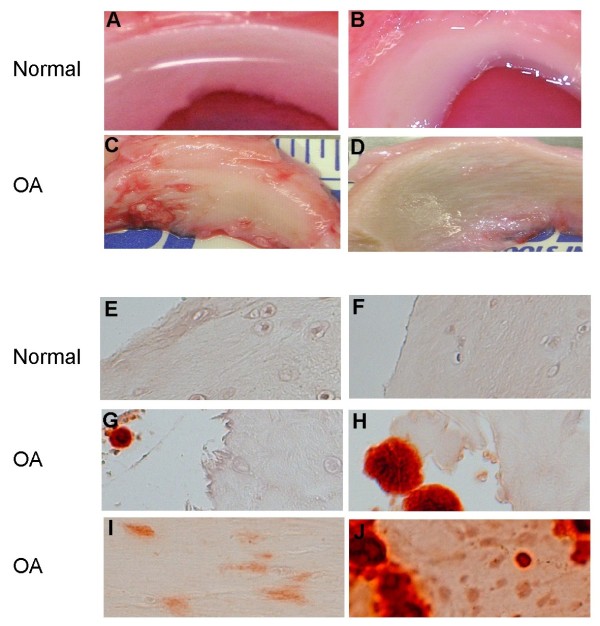
Introduction: Calcium crystals exist in the knee joint fluid of up to 65% of osteoarthritis (OA) patients and the presence of these calcium crystals correlates with the radiographic evidence of hyaline cartilaginous degeneration. This study sought to examine calcium deposition in OA meniscus and to investigate OA meniscal cell-mediated calcium deposition. The hypothesis was that OA meniscal cells may play a role in pathological meniscal calcification.
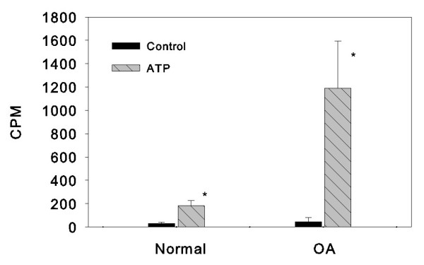
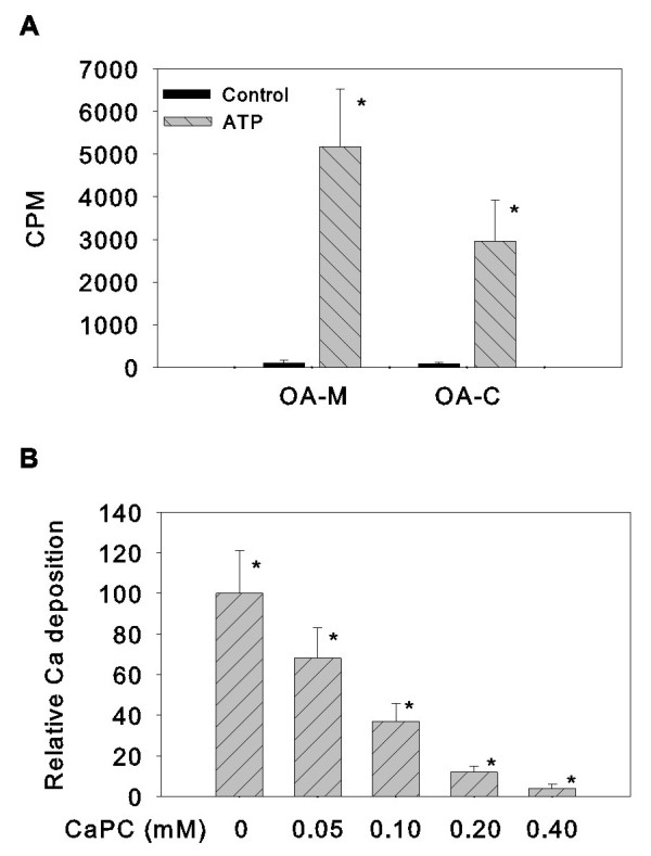
Methods: Studies were approved by our human subjects Institutional Review Board. Menisci were collected during joint replacement surgeries for OA patients and during limb amputation surgeries for osteosarcoma patients. Calcium deposits in menisci were examined by alizarin red staining. Expression of genes involved in biomineralization in OA meniscal cells was examined by microarray and real-time RT-PCR. Cell-mediated calcium deposition in monolayer culture of meniscal cells was examined using an ATP-induced (45)calcium deposition assay.
Results: Calcium depositions were detected in OA menisci but not in normal menisci. The expression of several genes involved in biomineralization including ENPP1 and ANKH was upregulated in OA meniscal cells. Consistently, ATP-induced calcium deposition in the monolayer culture of OA meniscal cells was much higher than that in the monolayer culture of control meniscal cells.
Conclusions: Calcium deposition is common in OA menisci. OA meniscal cells calcify more readily than normal meniscal cells. Pathological meniscal calcification, which may alter the biomechanical properties of the knee meniscus, is potentially an important contributory factor to OA.
Figures
Comment in
-
The meniscus, calcification and osteoarthritis: a pathologic team.Arthritis Res Ther. 2010;12(3):116. doi: 10.1186/ar2993. Epub 2010 May 20. Arthritis Res Ther. 2010. PMID: 20500910 Free PMC article.
References
-
- Samuels J, Krasnokutsky S, Abramson SB. Osteoarthritis: a tale of three tissues. Bull NYU Hosp Jt Dis. 2008;66:244–250. - PubMed
-
- Kato H, Matsumine A, Wakabayashi T, Hasegawa M, Sudo A, Shintani K, Fukuda A, Kato K, Ide N, Orita S, Hasegawa T, Matsumura C, Furukawa M, Tasaki T, Sonoda H, Uchida A. Large-scale gene expression profiles, differentially represented in osteoarthritic synovium of the knee joint using cDNA microarray technology. Biomarkers. 2007;12:384–402. doi: 10.1080/13547500601162482. - DOI - PubMed
Publication types
MeSH terms
Substances
LinkOut - more resources
Full Text Sources
Other Literature Sources
Miscellaneous
