. 2010 Mar 30:11:17.
doi: 10.1186/1471-2172-11-17.

Structural and functional studies of STAT1 from Atlantic salmon (Salmo salar)

Affiliations

Structural and functional studies of STAT1 from Atlantic salmon (Salmo salar)

Astrid Skjesol et al. BMC Immunol. .

Abstract

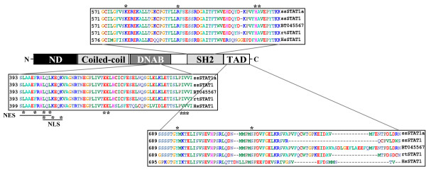
Background: Type I and type II interferons (IFNs) exert their effects mainly through the JAK/STAT pathway, which is presently best described in mammals. STAT1 is involved in signaling pathways induced by both types of IFNs. It has a domain-like structure including an amino-terminus that stabilizes interaction between STAT dimers in a promoter-binding situation, a coiled coil domain facilitating interactions to other proteins, a central DNA-binding domain, a SH2 domain responsible for dimerization of phosphorylated STATs and conserved phosphorylation sites within the carboxy terminus. The latter is also the transcriptional activation domain.

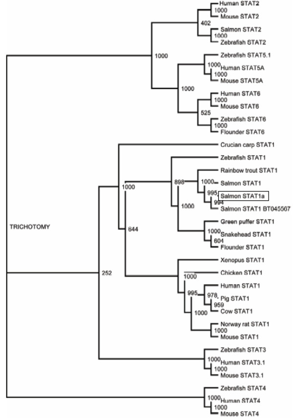
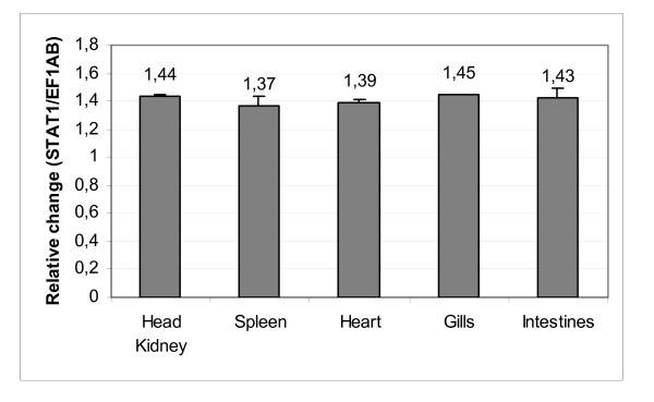
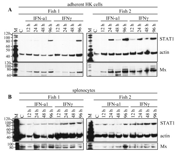
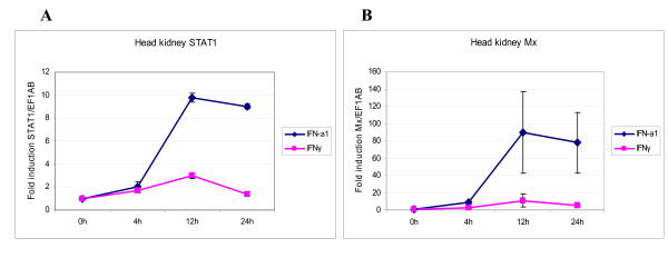
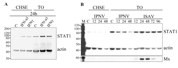
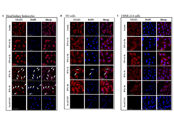
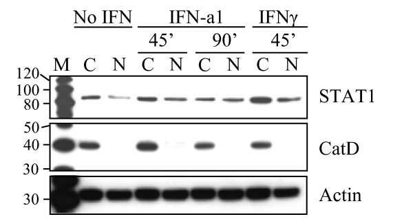
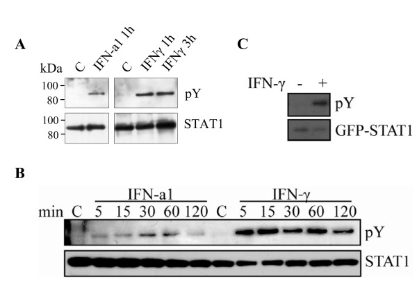
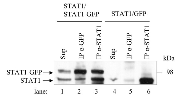
Results: A salmon (Salmo salar) STAT1 homologue, named ssSTAT1a, has been identified and was shown to be ubiquitously expressed in various cells and tissues. The ssSTAT1a had a domain-like structure with functional motifs that are similar to higher vertebrates. Endogenous STAT1 was shown to be phosphorylated at tyrosine residues both in salmon leukocytes and in TO cells treated with recombinant type I and type II IFNs. Also ectopically expressed ssSTAT1 was phosphorylated in salmon cells upon in vitro stimulation by the IFNs, confirming that the cloned gene was recognized by upstream tyrosine kinases. Treatment with IFNs led to nuclear translocation of STAT1 within one hour. The ability of salmon STAT1 to dimerize was also shown.

Conclusions: The structural and functional properties of salmon STAT1 resemble the properties of mammalian STAT1.


Figures

Figure 1
Figure 1
Salmon STAT1 protein harbors conserved domains and sequences. The NCBI conserved domains database and ClustalW alignment was combined to depict the schematic presentation of STAT1. Abbreviations: ND = amino-terminal domain, DNAD = DNA-binding domain, SH2 = Src Homology 2 domain, TAD = transcriptional activation domain, NES = nuclear export signal, NLS = nuclear localization signal, ss = Atlantic salmon, rt = rainbow trout, hs = human. Asterisks indicate conserved residues with importance for the functional activity within the domains.
Figure 2
Figure 2
STAT1 phylogeny. An un-rooted phylogenetic tree of STAT1 and other STAT proteins based on sequences aligned by ClustalW was constructed using the neighbor-joining algorithm. Bootstrap value = 1000.
Figure 3
Figure 3
STAT1 mRNA is evenly distributed in salmon tissues. STAT1 mRNA levels in the salmon tissues head kidney, spleen, heart, gills and intestines was detected by qPCR analysis. The expression levels are presented as change in expression relative to EF1AB. Samples from 10 fish were tested.
Figure 4
Figure 4
SDS-PAGE followed by Western blot showing expression of STAT1 protein in (A) adherent head kidney leukocytes and (B) splenocytes from two individuals upon stimulation with IFN. The samples were harvested 12, 24, 48, and 96 h after stimulation with IFN-a1 (10 U/mL) and IFNγ (200 ng/mL). The unstimulated control, C, was harvested at the 48 h time-point. STAT1 protein was detected simultaneously with actin which was used as a loading control. The membranes were stripped and reprobed with an anti-Mx-antibody as a control for the IFN-activity. M = MagicMark molecular weight marker.
Figure 5
Figure 5
QPCR showing expression of STAT1 and Mx mRNA in IFN-stimulated adherent head kidney leukocytes. The cells were stimulated with 10 U/mL of recombinant Atlantic salmon IFN-a1 or 200 ng/mL of recombinant rainbow trout IFNγ. The mRNA levels were normalized against EF1AB. (A) STAT1 mRNA expression after 4, 12 and 24 h of stimulation. (B) Mx mRNA expression after 4, 12 and 24 h of stimulation. The results are an average of samples from three fish and presented as fold increase relative to unstimulated head kidney cells. Experiments were performed twice with reproducible results. Error bars show standard deviation.
Figure 6
Figure 6
SDS-PAGE followed by Western blot showing expression of STAT1 protein in salmonid cell-lines. Antibodies against salmon STAT1 (α-STAT1 1:2,000), actin (α-actin, 1:1,000) and Mx (α-Mx, 1:1,000) were used. (A) CHSE-214 and TO cells were harvested 24 h after stimulation with IFN-a1 (10 U/mL) and IFNγ (200 ng/mL) along with an unstimulated control, C. (B) CHSE-214 and TO cells were infected with IPNV (MOI = 4) and harvested at 12, 24 and 48 h p.i. TO cells were also infected with ISAV (MOI = 4) and harvested at 12, 24, 48, 72 and 96 h p.i. Actin was used as a loading control and Mx protein detected after the membrane was stripped and reprobed. M = MagicMark molecular weight marker.
Figure 7
Figure 7
Subcellular localization of STAT1 in different cell-types after stimulation with IFN-a1 and IFNγ. The cells were treated with IFN-a1 (10 U/mL) or IFNγ (200 ng/mL) for 1 or 4 h or left unstimulated before fixed in 4% paraformaldehyde and stained for STAT1 (red). Nuclei were stained with DAPI (blue). (A) Salmon adherent head kidney leukocytes. Translocation of STAT1 to the nucleus took place at 1 h and at 4 h after stimulation with IFNγ as indicated by arrows. (B) TO cells. Translocation of STAT1 to the nucleus took place about 1 h after stimulation with IFNγ as indicated by arrows. (C) CHSE-214 cells. No translocation of STAT1 to the nucleus was observed. A control with no α-STAT1 is included for each of the cell types.
Figure 8
Figure 8
Nuclear localization of STAT1 in TO cells after stimulation with IFN-a1 and IFNγ. The cells were treated with IFN-a1 (10 U/mL) for 45 or 90 min or IFNγ (200 ng/mL) for 45 min or left unstimulated. Nuclear extracts (N) and cytoplasmic fractions (C) were separated using NE-PER Nuclear and Cytoplasmic Extraction Reagents (Thermo scientific). The extracts were subjected to SDS-PAGE and Western blotting. STAT1 was detected in both N and C fractions, but lesser STAT1 was observed in the nuclear fraction of the unstimulated cells. An antibody directed against a strictly lysosomal protein, Cathepsin D (CatD) was used as a control for cytoplasmic localization. After stripping the membrane, an actin antibody was applied as a loading control. M = MagicMark molecular weight marker.
Figure 9
Figure 9
STAT1 is phosphorylated in response to IFN-a1 and IFNγ. Cells were either treated with IFN-a1 (10 U/ml) or IFN-γ (200 ng/ml) or left untreated. Cells were harvested at indicated time points and endogenous STAT1 were immunoprecipitated with α-STAT1 from the whole cell extracts. Tyrosine phosphorylated STAT1 was detected by immunoblotting using anti-phosphotyrosine antibody (pY, upper panel). The total amount of immunoprecipitated STAT1 was detected with α-STAT1 antibody (lower panel). (A) Head kidney leukocytes. (B) TO cells. (C) TO cells were transfected with a GFP-ssSTAT1a construct. After 48 h, the cells were treated with IFNγ (200 ng/ml) for 30 min, or left untreated. Cells were lysed and GFP-tagged proteins were immunoprecipitated with α-GFP. Phosphorylated GFP-STAT1 was detected by immunoblotting using anti-phosphotyrosine antibody (upper panel). The total amount of immunoprecipitated GFP-STAT1 was detected with α-GFP antibody (lower panel).
Figure 10
Figure 10
Co-IP analyses of the ssSTAT1a - ssSTAT1a interaction. ssSTAT1a was co-expressed with GFP-ssSTAT1a or pEXP-GFP (negative control) in HEK-293 cells and the lysed cells subjected to IP with a STAT1 antibody (α-STAT1) or a GFP antibody (α-GFP). Samples were analyzed along with the total cell lysate (sup) by SDS-PAGE followed by Western blot using α-STAT1.

Similar articles

Cited by

References

    1. Samuel CE. Antiviral actions of interferons. Clin Microbiol Rev. 2001;14(4):778–809. doi: 10.1128/CMR.14.4.778-809.2001. - DOI - PMC - PubMed
    1. Isaacs A, Lindenmann J. Virus Interference .1. The Interferon. Proc R Soc Lond Ser B-Biol Sci. 1957;147(927):258–267. doi: 10.1098/rspb.1957.0048. - DOI - PubMed
    1. Platanias LC. Mechanisms of type-I- and type-II-interferon-mediated signalling. Nat Rev Immunol. 2005;5(5):375–386. doi: 10.1038/nri1604. - DOI - PubMed
    1. Aaronson DS, Horvath CM. A Road Map for Those Who Don't Know JAK-STAT. Science. 2002;296(5573):1653–1655. doi: 10.1126/science.1071545. - DOI - PubMed
    1. Brierley MM, Fish EN. Stats: Multifaceted regulators of transcription. Journal of Interferon and Cytokine Research. 2005;25(12):733–744. doi: 10.1089/jir.2005.25.733. - DOI - PubMed

Publication types

LinkOut - more resources