

Folliculostellate cells in pituitary pars distalis of male viscacha: immunohistochemical, morphometric and ultrastructural study
- PMID: 20353904
- PMCID: PMC3167288
- DOI: 10.4081/ejh.2010.e1
Folliculostellate cells in pituitary pars distalis of male viscacha: immunohistochemical, morphometric and ultrastructural study
Abstract
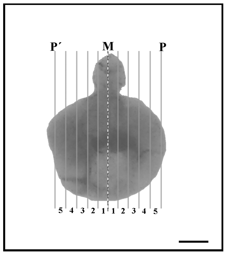
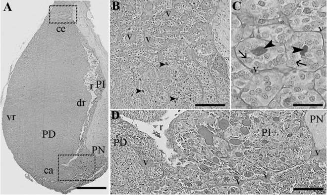
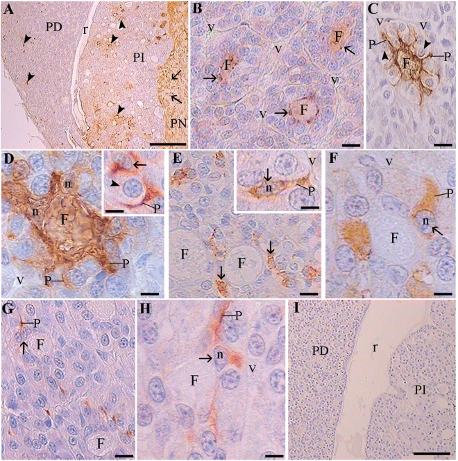
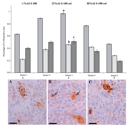
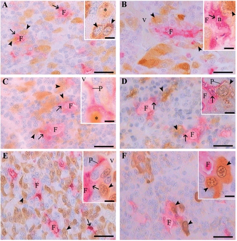
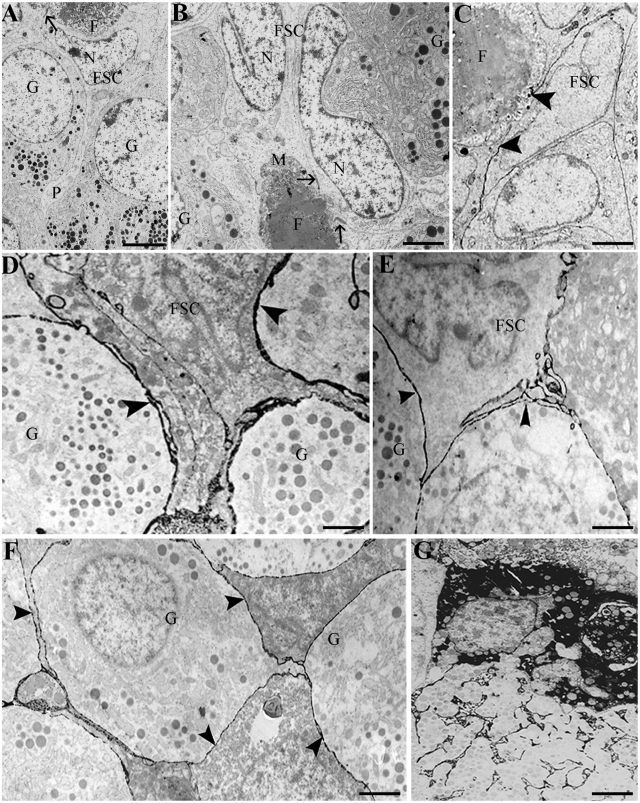
Folliculostellate cells (FSC) have been reported in pituitary of several mammalian species. FSC morphology and secreted substances have been instrumental to the understanding of their function. The purpose of this work was to perform an immunohistochemical, morphometric and ultrastructural study of the pituitary pars distalis FSC in adult male viscacha and to analyze their relation with hormone secreting cells. Immunohistoche-mistry and image analysis were carried out in different sectors of the gland, from the middle (sector 1) to the glandular periphery (sector 5). Transmission electron microscopy with lanthanum as electrodense tracer was used. FSC formed follicles with PAS-positive colloid inside. They expressed S-100 protein mainly in both nucleus and cytoplasm. FSC were stellate-like in shape and exhibited short cytoplasmic processes that contacted with blood vessels and endocrine cells. In addition, some follicular colloids were immunostained with anti-S-100 protein. A few FSC were immunostained with anti-glial fibrillary acidic protein (GFAP) and anti-vimentin. The morphometric parameters analyzed (percentages of S-100-positive total, cellular and colloidal areas) increased from sector 1 to sector 3 and then decreased to sector 5. Hormone secreting cells, mainly lactotrophs, gonadotrophs and corticotrophs were associated with FSC and follicles. The ultrastructural study demonstrated that FSC developed junctional complexes and desmosomes between their lateral membranes. Lanthanum freely penetrated the spaces between granulated cells and FSC, but did not penetrate into the follicular lumen.
In conclusion: 1) the differential expression of S-100 protein, GFAP and vimentin may indicate different physiological stages of FSC; 2) the expression of these proteins suggests a neuroectodermic origin of these cells; 3) FSC spatial distribution, association with endocrine cells, and the generation of an intercellular communication network suggest that FSC are involved in the pituitary pars distalis paracrine regulation of the viscacha.
Figures
References
-
- Marin F, Stefaneanu L, Kovacs K. Folliculostellate cells of the pituitary. Endocr Pathol. 1991;2:180–92. - PubMed
-
- Allaerts W, Vankelecom H. History and perspectives of pituitary folliculo-stellate cell research. Eur J Endocrinol. 2005;153:1–12. - PubMed
-
- Luziga C, Yoshimi Y, Yoichiro H, et al. Phagocytotic removal of apoptotic endocrine cells by folliculostellate cells and its functional implications in clusterin accumulation in pituitary colloids in helmeted guinea fowl (Numida meleagris) Acta Histochem. 2006;108:69–80. - PubMed
-
- Luziga C, Kipanyula MJ, Mbassa G, Koichi M. Colloid in the anterior pituitary of helmet guinea fowl (Numida meleagris galeata): morphometric analysis and pattern of occurrence in relation to apoptosis. Vet Res Commun. 2009;33:681–91. - PubMed
-
- Devnath S, Inoue K. An insight to pituitary folliculo-stellate cells. J Neuroendocrinol. 2008;20:687–91. - PubMed
Publication types
MeSH terms
Substances
LinkOut - more resources
Full Text Sources
Miscellaneous
