

Distribution of NADPH-diaphorase and AChE activity in the anterior leaflet of rat mitral valve
- PMID: 20353912
- PMCID: PMC3167287
- DOI: 10.4081/ejh.2010.e5
Distribution of NADPH-diaphorase and AChE activity in the anterior leaflet of rat mitral valve
Abstract
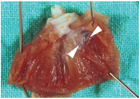
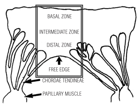
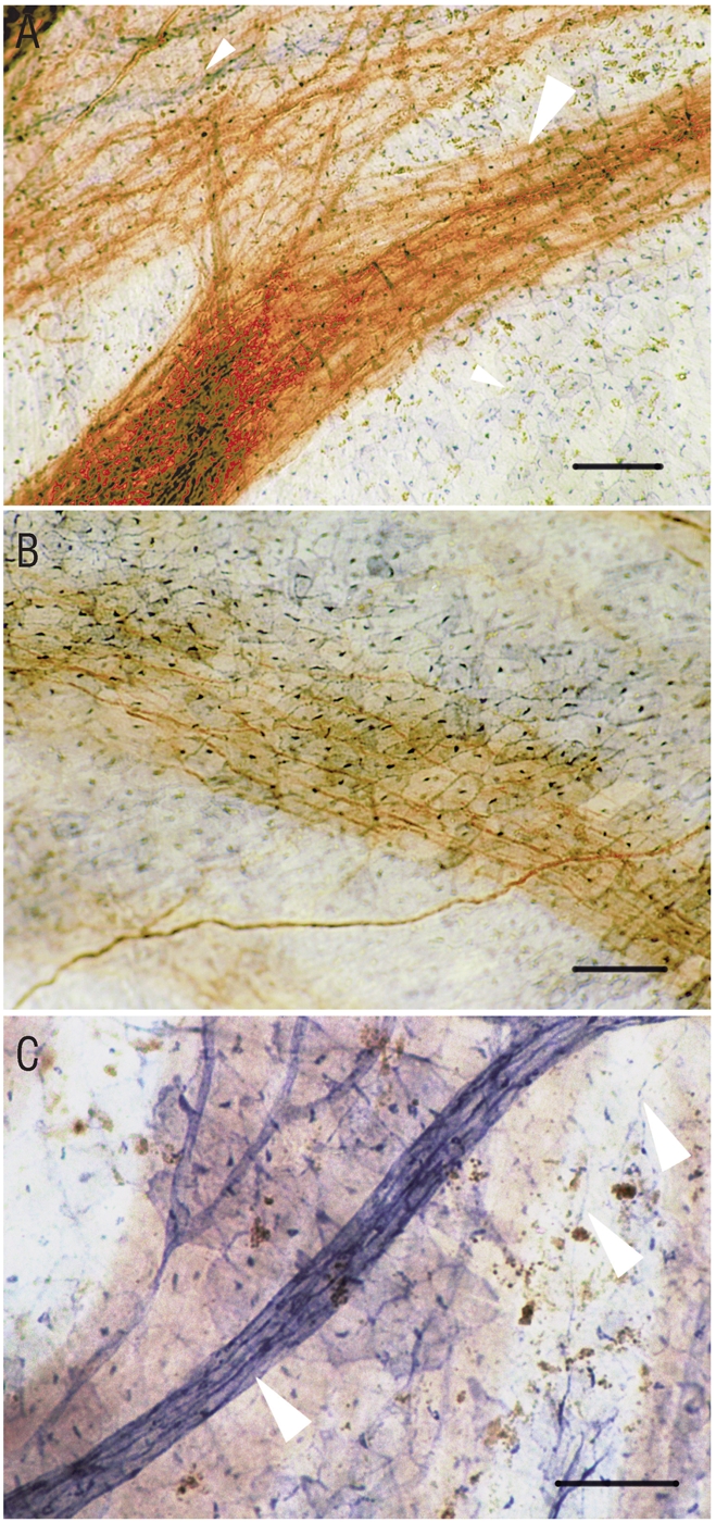
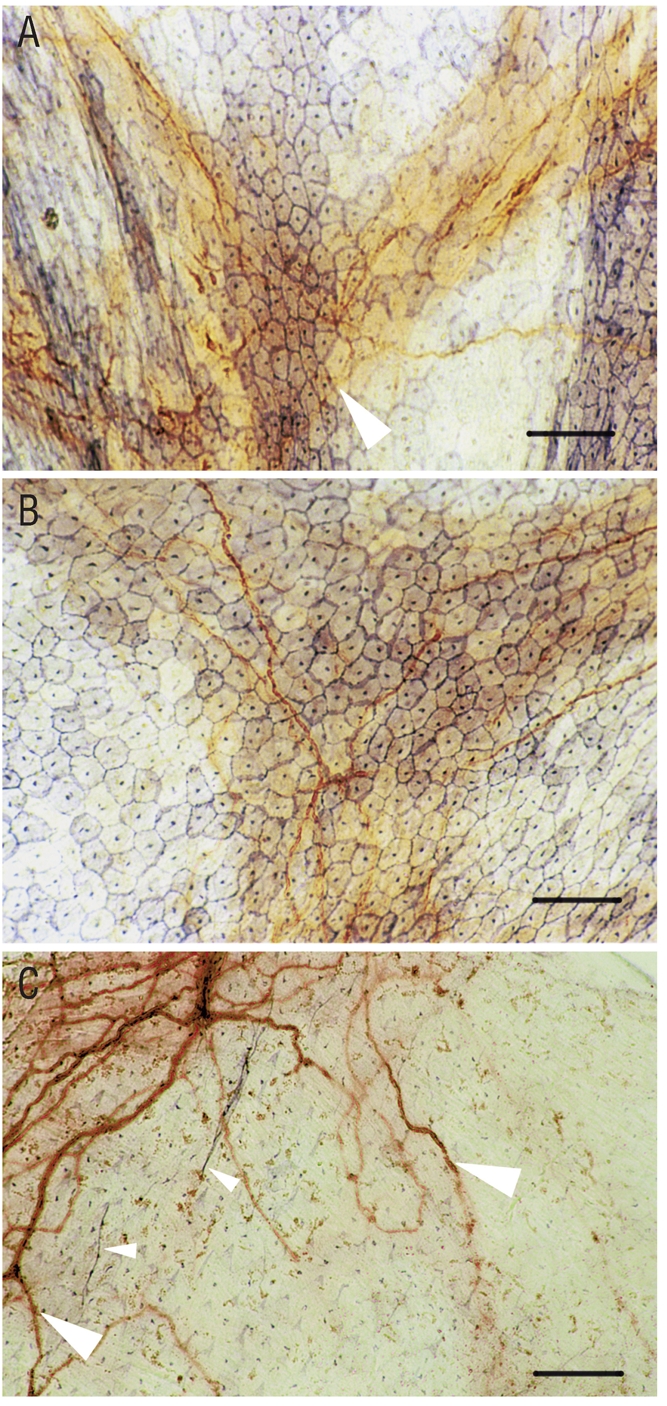
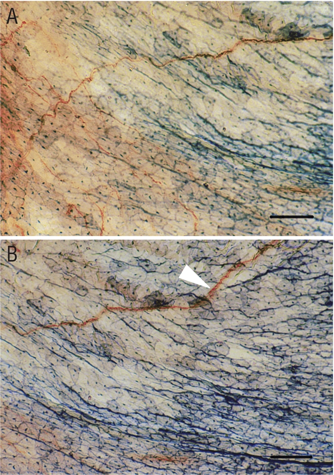
The mitral valve, as an active flap, forms the major part of the left ventricular inflow tract and therefore plays an important function in many aspects of left ventricular performance. The anterior leaflet of this valve is the largest and most ventrally placed of two leaflets that come together during ventricular systole to close the left atrioventricular orifice. Various neurotransmitters are responsible for different functions including controlling valve movement, inhibiting or causing the failure of impulse conduction in the valve and the sensation of pain. Nitric oxide acts as a gaseous free radical neurotransmitter, neuromediator and effective cardiovascular modulator. Acetyl-choline is known to function as a typical neurotransmitter. Histochemical methods for detection of nicotinamide adenine dinucleotide phosphate diaphorase (NADPH-d), as an indirect nitric oxide-synthase marker, and method for detection of acetylcholinesterase (AChE) were used. Both methods were performed on the same valve sample. A widespread distribution of nerve fibres was observed in the anterior leaflet of the mitral valve. The fine NADPH-d positive (nitrergic) nerve fibres were identified in all zones of valve leaflet. AChE positive (cholinergic) nerve fibres were identified forming dense network and fibres organized in stripes. Endocardial cells and vessels manifested heavy NADPH-d activity. Our observations suggest a different arrangement of nitrergic and cholinergic nerve fibres in the anterior leaflet of the mitral valve. The presence of nitrergic and cholinergic activity confirms the involvement of both neurotransmitters in nerve plexuses and other structures of mitral valve.
Figures
Similar articles
-
Postnatal development of nitrergic and cholinergic structures in rat spinal cord.Arch Ital Biol. 2011 Sep;149(3):293-302. doi: 10.4449/aib.v149i3.1219. Arch Ital Biol. 2011. PMID: 22028090
-
Increased NADPH-diaphorase activity in canine myxomatous mitral valve leaflets.J Comp Pathol. 2003 Aug-Oct;129(2-3):120-30. doi: 10.1016/s0021-9975(03)00019-7. J Comp Pathol. 2003. PMID: 12921717
-
Histochemical study of innervation and NADPH-D activity of the thymus.Anat Histol Embryol. 2003 Aug;32(4):233-5. doi: 10.1046/j.1439-0264.2003.00470.x. Anat Histol Embryol. 2003. PMID: 12919075
-
[Endogenously formed nitric oxide in nasal mucosa of the human: detection by nicotinamide-adenine dinucleotide phosphate diaphorase (NADPH-d) histochemistry].Laryngorhinootologie. 1996 Oct;75(10):584-9. doi: 10.1055/s-2007-997639. Laryngorhinootologie. 1996. PMID: 9035661 German.
-
The effects on cordal and leaflet stiffness of severe apical, posterior, and outward papillary displacement in advanced ventricular mechanism heart failure and mitral insufficiency.J Heart Valve Dis. 2011 Nov;20(6):608-18. J Heart Valve Dis. 2011. PMID: 22655489 Review.
Cited by
-
Histochemistry through the years, browsing a long-established journal: novelties in traditional subjects.Eur J Histochem. 2010 Dec 16;54(4):e51. doi: 10.4081/ejh.2010.e51. Eur J Histochem. 2010. PMID: 21263750 Free PMC article.
-
NADPH-diaphorase expression in the rat jejunum after intestinal ischemia/reperfusion.Eur J Histochem. 2011;55(3):e23. doi: 10.4081/ejh.2011.e23. Epub 2011 Aug 27. Eur J Histochem. 2011. PMID: 22073370 Free PMC article.
-
On the future contents of a small journal of histochemistry.Eur J Histochem. 2012 Dec 10;56(4):e51. doi: 10.4081/ejh.2012.e51. Eur J Histochem. 2012. PMID: 23361247 Free PMC article.
-
NADPH-diaphorase expression in the meibomian glands of rat palpebra in postnatal development.Eur J Histochem. 2010 Nov 23;54(4):e47. doi: 10.4081/ejh.2010.e47. Eur J Histochem. 2010. PMID: 21263746 Free PMC article.
-
Effect of retinoic acid on the nitrergic innervation of meibomian glands in rats.Eur J Histochem. 2012 Nov 30;56(4):e50. doi: 10.4081/ejh.2012.e50. Eur J Histochem. 2012. PMID: 23361246 Free PMC article.
References
-
- Jew JY, Fink CA, Williams TH. Tyrosine hydroxylase-and nitric oxide synthase-immunoreactive nerve fibers in mitral valve of young adult and aged Fischer 344 rats. J Autonom Nerv Syst. 1996;58:35–43. - PubMed
-
- Fann JI, Ingels NB, Miller DC. Pathophysiology of mitral valve disease. In: Cohn LH, Edmunds LH Jr., editors. Cardiac Surgery in the Adult. McGraw-Hill; New York: 2003. pp. 901–931.
-
- Kachlik D, Baca V, Bozdechova I, et al. Anatomical terminology and nomenclature: past, presence and highlights. Surg Rad Anat. 2008;30:459–66. - PubMed
-
- Yacoub MH, Cohn LH. Novel approaches to cardiac valve repair: From structure to function: Part I. Circulation. 2004;109:942–50. - PubMed
Publication types
MeSH terms
Substances
LinkOut - more resources
Full Text Sources
