. 2010 Jun 4;285(23):17564-73.
doi: 10.1074/jbc.M110.111641. Epub 2010 Mar 30.

Cis and trans actions of the cholinesterase-like domain within the thyroglobulin dimer

Affiliations

Cis and trans actions of the cholinesterase-like domain within the thyroglobulin dimer

Xiaofan Wang et al. J Biol Chem. .

Abstract

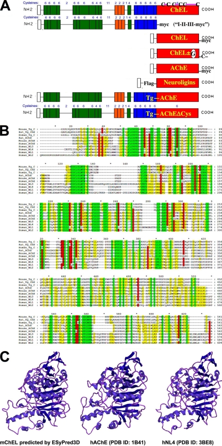
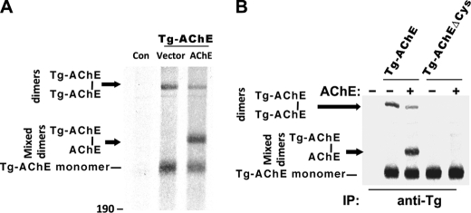
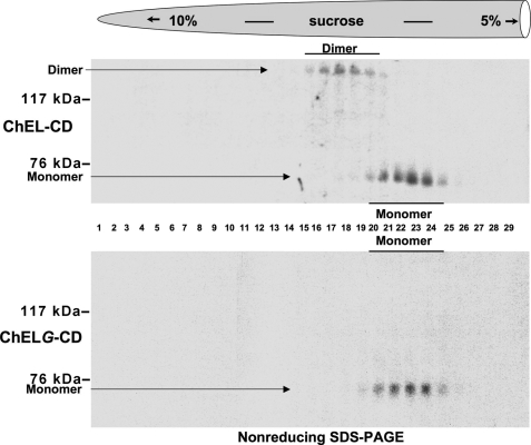
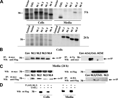
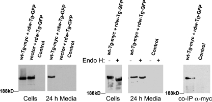
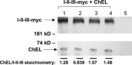
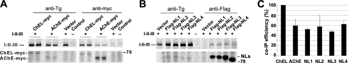
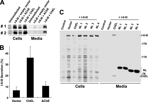
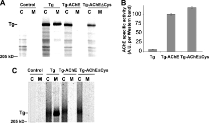
Thyroglobulin (Tg, precursor for thyroid hormone synthesis) is a large secreted glycoprotein composed of upstream regions I-II-III, followed by the approximately 570 residue cholinesterase-like (ChEL) domain. ChEL has two identified functions: 1) homodimerization, and 2) binding to I-II-III that facilitates I-II-III oxidative maturation required for intracellular protein transport. Like its homologs in the acetylcholinesterase (AChE) family, ChEL possesses two carboxyl-terminal alpha-helices. We find that a Tg-AChE chimera (swapping AChE in place of ChEL) allows for dimerization with monomeric AChE, proving exposure of the carboxyl-terminal helices within the larger context of Tg. Further, we establish that perturbing trans-helical interaction blocks homodimerization of the Tg ChEL domain. Additionally, ChEL can associate with neuroligins (a related family of cholinesterase-like proteins), demonstrating potential for Tg cross-dimerization between non-identical partners. Indeed, when mutant rdw-Tg (Tg-G2298R, defective for protein secretion) is co-expressed with wild-type Tg, the two proteins cross-dimerize and secretion of rdw-Tg is partially restored. Moreover, we find that AChE and soluble neuroligins also can bind to the upstream Tg regions I-II-III; however, they cannot rescue secretion, because they cannot facilitate oxidative maturation of I-II-III. These data suggest that specific properties of distinct Tg ChEL mutants may result in distinct patterns of Tg monomer folding, cross-dimerization with wild-type Tg, and variable secretion behavior in heterozygous patients.


Figures

FIGURE 1.
FIGURE 1.
Homologs of the Tg ChEL domain. A, constructs used in this study, in relation to the domain structure of Tg shown schematically. Top to bottom: the concluding ChEL domain of wild-type Tg (uppermost) contains six Cys residues, and based on conservation of structure with AChE, the mature mouse Tg ChEL domain is predicted to have three disulfide bonds engaging Cys2243–Cys2260, Cys2421–Cys2432, and Cys2570–Cys2694. Not shown in the schematic are engineered Tg variants, including wild-type Tg bearing a carboxyl-terminal Myc epitope tag, and rdw-Tg (Tg-G2298R) bearing carboxyl-terminal GFP (see Fig. 5). A Myc-tagged Tg construct lacking the ChEL domain is called “I-II-III-Myc.” Next shown is secretory ChEL bearing a carboxyl-terminal Myc tag. Thereafter, secretory ChEL bearing an extra unpaired carboxyl-terminal Cys residue for covalent dimerization (ChEL-CD) was engineered to either include an Asn-linked glycosylation site (called “ChELG-CD”) or lack that site. Next, AChE bearing a carboxyl-terminal Myc tag is shown; note that AChE already contains a naturally occurring seventh (extra, unpaired) Cys residue at its carboxyl terminus. Next, secretory (FLAG-tagged) neuroligins 1, 2, 3, or 4 are indicated. Finally, a Tg-AChE chimera, which already contains a natural seventh (extra, unpaired) Cys residue at its carboxyl terminus (14), is shown; an identical Tg-AChE chimera lacking the carboxyl-terminal Cys residue (called Tg-AChEΔCys) was also prepared. B, multiple sequence alignment with mouse Tg, listed in order of greatest homology, of the primary structures derived from rat Tg, human Tg, rat AChE, mouse AChE, human AChE, and human neuroligins (NL) 3, 4, 1, and 2, respectively. Areas of perfect homology are colored green; 80 and 60% conservation are colored red and yellow, respectively. C, a comparison of the known structure of human AChE (PDB ID: 1B41) and human NL4 (PDB ID: 3BE8) with the three-dimensional structure of mouse ChEL predicted by the ESyPred3D structural prediction program.
FIGURE 2.
FIGURE 2.
Availability of AChE dimerization helices within the context of a Tg-AChE chimera. A, 293 cells were transiently co-transfected with empty vector + vector (“Con”) or with plasmids expressing Tg-AChE (14) plus either vector or human AChE. The cells were metabolically labeled with 35S-amino acids and chased for 5 h in the presence of 5 μg/ml brefeldin A. The cell lysates were immunoprecipitated with anti-Tg and analyzed by non-reducing SDS-4%-PAGE. Monomeric Tg-AChE migrated with an apparent molecular mass of ∼300 kDa; the covalent Tg-AChE homodimer migrated at ∼600 kDa; the position of mixed Tg-AChE–AChE covalent dimers is shown. B, the Tg-AChE chimera construct was mutagenized to substitute its extra unpaired Cys residue with Ser near the carboxyl terminus to create Tg-AChEΔCys. Pulse-labeling and chase of co-transfected 293 cells included either Tg-AChE or Tg-AChEΔCys with either empty vector (−) or a plasmid to co-express AChE (+), as indicated. Samples were analyzed as in panel A.
FIGURE 3.
FIGURE 3.
Tail-to-tail ChEL homodimerization. Upper gradient: 293 cells transiently expressing secretory ChEL-CD were pulse-labeled with 35S-amino acids and chased, and the secretion was resolved on 5–10% linear sucrose velocity gradients as described under “Experimental Procedures.” Fractions collected from the bottom (left side) were immunoprecipitated with anti-Tg antiserum and analyzed by non-reducing SDS-PAGE and fluorography. Monomers sedimented between fractions 20 and 25; dimers sedimented between fractions 15 and 20. Monomeric ChEL-CD migrated by SDS-PAGE with an apparent molecular mass of ∼70 kDa; covalent ChEL-CD dimer migrated with an apparent molecular mass of ∼140 kDa. Lower gradient: 293 cells transiently expressing the secretory ChELG-CD mutant were analyzed as above. None of the ChELG-CD contained an intermolecular covalent bond; all molecules migrated to the monomer peak in the gradient.
FIGURE 4.
FIGURE 4.
Cross-dimerization with ChEL. A, 293 cells were transiently transfected with empty vector or plasmids to express secretory ChEL-Myc (ChEL) or secretory FLAG-NL1, -NL2, -NL3, or -NL4. Transfected cells were pulse-labeled for 30 min with 35S-amino acids and chased for either 5 h (upper panel, a time when ChEL secretion has gone to completion) or 24 h (lower panel, a time when neuroligin secretion has gone to completion). The cells were lysed, and both cells and media were immunoprecipitated with anti-Myc (for ChEL) and anti-FLAG (for neuroligins). B, the left panel shows 293 cells co-transfected with plasmids expressing secretory ChEL-Myc and either empty vector (Con) or secretory FLAG-NL1, -NL2, -NL3, or -NL4. Cell lysates were immunoprecipitated (IP) with α-Myc antibody, resolved by SDS-PAGE and immunoblotted (WB) with anti-FLAG antibody. The right panel shows 293 cells co-transfected with plasmids expressing secretory ChEL and either empty vector (Con) or AChE-Myc (AChE). The cells were lysed in the presence of 1 mm dithiobis(succinimidyl propionate) cross-linker, immunoprecipitated (IP) with α-Tg antibody, resolved by SDS-PAGE and immunoblotted (WB) with anti-Myc antibody. C, the left panel shows 293 cells co-transfected as in the left panel of B, or transfected with empty vector (Con). The right panel shows 293 cells co-transfected with plasmids expressing secretory ChEL-HA plus secretory FLAG-NL3, or secretory FLAG-NL3 alone, or transfected with empty vector (Con). Media were collected from transfected cells for 24 h; the upper left panel shows the comparable expression of secretory ChEL partner in each sample; the upper right panel shows comparable expression of the secretory NL3 partner. Media were immunoprecipitated (IP) with α-Myc (lower left) or anti-HA (lower right); the immunoprecipitates were resolved by SDS-PAGE and immunoblotted (WB) with anti-FLAG antibodies. D, cells were transfected to either singly express secretory FLAG-NL3, singly express secretory ChEL-HA, or co-express both constructs, or neither. After 24 h, the cells were lysed and the media were collected; samples were analyzed by Western blotting with anti-FLAG antibodies.
FIGURE 5.
FIGURE 5.
Secretion rescue of mutant rdw-Tg by co-expressed wild-type Tg. Side-by-side, identical wells of 293 cells were co-transfected with plasmids encoding empty vector plus wild-type Tg-Myc (wt-Tg-Myc) and rdw-Tg-GFP such that total plasmid DNA and rdw-Tg-GFP DNA was held constant in all samples (wt:rdw plasmid ratio 6:1); Control lanes were transfected only with empty vector. At 48 h post-transfection, the medium was changed, and cells were cultured for an additional 24 h in complete growth medium. The cells were lysed, and 3.33% of each cell lysate and 1% of each collection of medium were resolved by SDS-PAGE and analyzed by immunoblotting with anti-GFP antibody. Left panels: direct immunoblotting of lysates and media. Middle panels: endoglycosidase H (Endo H) digestion of the samples indicated prior to immunoblotting. Note that the small mobility shift observed after endo H digestion of the medium is distinct from the larger shift seen for intracellular Tg, indicating Golgi sugar modification of Asn-linked glycans on all secreted rdw-Tg-GFP molecules (although some sugar chains on each molecule remain endo H-sensitive). Right panel: media from co-transfected or control cells were immunoprecipitated with α-Myc antibody, and co-precipitated rdw-Tg-GFP (co-IP) was analyzed by immunoblotting as in the other panels.
FIGURE 6.
FIGURE 6.
Equimolar ChEL:I-II-III binding stoichiometry. 293 cells were co-transfected to express I-II-III-Myc and secretory ChEL. In quadruplicate, transfected cells were metabolically labeled in complete growth medium (containing serum) plus 1 mCi/ml pure [35S]cysteine at fixed specific radioactivity. One day later, the medium was removed and replaced with fresh labeling medium for a further day before collection of secretion with analysis by immunoprecipitation with anti-Myc antibody, SDS-PAGE, fluorography, and scanning densitometry of the relevant bands. The stoichiometry of co-precipitated ChEL:I-II-III was quantified by correcting for relative isotope abundance. (6:116 reflects the ratio of Cys residues present in each binding partner. For purposes of easy visualization we increased exposure of the ChEL bands shown in the figure.) From these measurements, the average molar ratio was 1.17 ± 0.28, i.e. an equimolar binding stoichiometry. Lane 5 shows results from untransfected control cells. Results shown here were repeated and confirmed in two independent experiments (not shown).
FIGURE 7.
FIGURE 7.
AChE and secretory neuroligin association with Tg regions I-II-III. A, 293 cells were co-transfected with plasmids expressing I-II-III (or a negative control marked with “−”) and either secretory ChEL-Myc or AChE-Myc. The cells were pulse-labeled with 35S-amino acids and chased for 6 h in the presence of brefeldin A (to block all protein export). At the conclusion of the experiment, the cells were lysed in a non-denaturing lysis buffer containing 1% Nonidet P-40 but lacking SDS, and then immunoprecipitated with either anti-Tg or anti-Myc antibodies for analysis by SDS-PAGE and fluorography. Anti-Tg antibodies directly immunoprecipitated I-II-III; anti-Myc antibodies could recover I-II-III only if co-immunoprecipitated with a Myc-tagged binding partner. Note (from Fig. 6) that the relative isotope abundance of Cys residues in one ChEL versus one I-II-III is a ratio of 6:116, explaining the relative intensities of ChEL and I-II-III. B, 293 cells were co-transfected with plasmids expressing I-II-III and either empty vector or secretory FLAG-NL1, -NL2, -NL3, or -NL4. At the 6-h chase time after pulse labeling, cell lysates were divided in half and immunoprecipitated with anti-Tg (first five lanes) or anti-FLAG (last five lanes) and analyzed by SDS-PAGE and fluorography. Anti-Tg antibodies directly immunoprecipitated I-II-III; anti-FLAG antibodies could recover I-II-III only if co-immunoprecipitated with a FLAG-tagged binding partner. C, quantitation of efficiency of co-immunoprecipitation of I-II-III with AChE or secretory neuroligins relative to co-precipitation obtained with ChEL (n = 3 experiments).
FIGURE 8.
FIGURE 8.
ChEL is unique in its ability to promote I-II-III secretion. A, in two independent transfections, 293 cells were co-transfected with the plasmids indicated at the top. The cells were pulse-labeled with 35S-amino acids for 30 min and chased for 4 h in complete medium. The cells were lysed and the media collected; both were immunoprecipitated with anti-Tg and analyzed by reducing SDS-PAGE and fluorography. B, from three experiments like those shown in panel A, secretion efficiency of I-II-III in the presence of empty vector, secretory ChEL, or AChE was quantified. C, 293 cells were co-transfected with plasmid expressing I-II-III and secretory FLAG-NL1, -NL2, -NL3, or -NL4 as indicated. Co-transfection with secretory ChEL was included as a positive control. The cells were pulse-labeled with 35S-amino acids for 30 min and chased for 4 h in complete medium. The cells were lysed, and the medium was collected; both were immunoprecipitated with a combination of anti-Tg plus anti-FLAG and analyzed by reducing SDS-PAGE and fluorography. As shown, secretory neuroligins could not rescue I-II-III secretion.
FIGURE 9.
FIGURE 9.
ChEL, but not AChE, promotes a stable, oxidatively mature state of regions I-II-III. A, 293 cells were transfected to express I-II-III alone (lanes 1–3) or co-express AChE (lanes 4–6) or secretory ChEL (lanes 7–9). The cells were pulse-labeled with 35S-amino acids for 30 min and chased in the presence of brefeldin A (5 μg/ml) in complete medium for the times indicated. At each chase time, the cells were lysed, immunoprecipitated with anti-Tg, and analyzed by non-reducing SDS-PAGE and fluorography. Bands A, B, and C have been described previously but are quantitatively minor (10); the positions of immature (D) and oxidatively mature (E) forms of I-II-III monomers are indicated. Untransfected cells (a negative control) are shown in lanes 10–12. B, 293 cells transfected to express I-II-III alone (middle lane) or co-express AChE-Myc (left) or secretory ChEL-KDEL (right), were pulse labeled as in A and chased (without brefeldin A) for 2 h. The cells were lysed and immunoprecipitated with anti-Tg; the immunoprecipitates were digested with PNGase F and analyzed by non-reducing SDS-PAGE and fluorography. The positions of immature (D) and mature (E) forms of I-II-III are indicated. C, 293 cells expressing I-II-III co-transfected with either empty vector (lanes 1–3) or secretory ChEL-Myc (lanes 4–6) or AChE-Myc (lanes 7–9). The cells were pulse-labeled with 35S-amino acids and chased as indicated. At each chase time, the cells were lysed, immunoprecipitated with anti-Myc, and analyzed by reducing SDS-PAGE (lower panel, the position of a 77-kDa molecular mass standard is shown on the left) to demonstrate relative production of ChEL-Myc (slower migrating) and AChE-Myc (faster migrating), or immunoprecipitated with anti-Tg and analyzed by non-reducing SDS-PAGE (upper panel, the position of a 190-kDa molecular mass standard is shown on the left) to examine the extent of oxidative maturation of Tg I-II-III. Note that, although AChE expression exceeds that of ChEL, stable, oxidatively mature I-II-III are recovered only in the presence of ChEL.
FIGURE 10.
FIGURE 10.
A Tg-AChE chimera exhibiting functional cholinesterase activity is nevertheless incompetent for secretion. A, 293 cells transfected to express either wild-type Tg (as a positive control), Tg-AChE, or Tg-AChEΔCys were pulse-labeled for 30 min and chased for 3.5 h. The media were collected, and the cells were lysed and immunoprecipitated with anti-Tg before analysis by reducing SDS-PAGE and fluorography. B, specific cholinesterase activity of Tg-AChE and Tg-AChEΔCys (normalized to Western blotting with anti-Tg). C, an independent experiment similar to panel A but analyzed by non-reducing SDS-PAGE and fluorography. Despite that AChE has a smaller molecular mass than ChEL, Tg-AChE and Tg-AChEΔCys run slower (higher), because they are oxidatively immature. Untransfected cells (Control) are shown in the first two lanes of panels A and C. The position of a 205-kDa molecular mass marker is indicated.

Similar articles

Cited by

References

    1. Di Jeso B., Arvan P. (2004) in The Thyroid (Braverman L. E., Utiger R. eds) 9th Ed., pp. 77–95, Lippincott Williams & Wilkins, Philadelphia
    1. Taurog A. (1999) Biochimie 81, 557–562 - PubMed
    1. Marriq C., Lejeune P. J., Venot N., Vinet L. (1991) Mol. Cell. Endocrinol. 81, 155–164 - PubMed
    1. Dunn A. D., Corsi C. M., Myers H. E., Dunn J. T. (1998) J. Biol. Chem. 273, 25223–25229 - PubMed
    1. Lamas L., Taurog A. (1977) Endocrinology 100, 1129–1136 - PubMed

Publication types