. 2010 Jun 4;285(23):17754-62.
doi: 10.1074/jbc.M109.090621. Epub 2010 Mar 30.

The human Cdc34 carboxyl terminus contains a non-covalent ubiquitin binding activity that contributes to SCF-dependent ubiquitination

Affiliations

The human Cdc34 carboxyl terminus contains a non-covalent ubiquitin binding activity that contributes to SCF-dependent ubiquitination

Yun-Seok Choi et al. J Biol Chem. .

Abstract

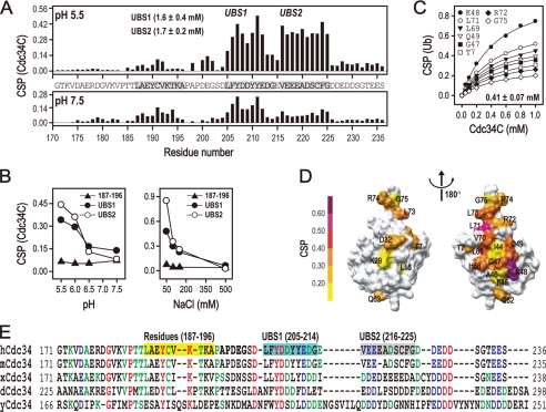
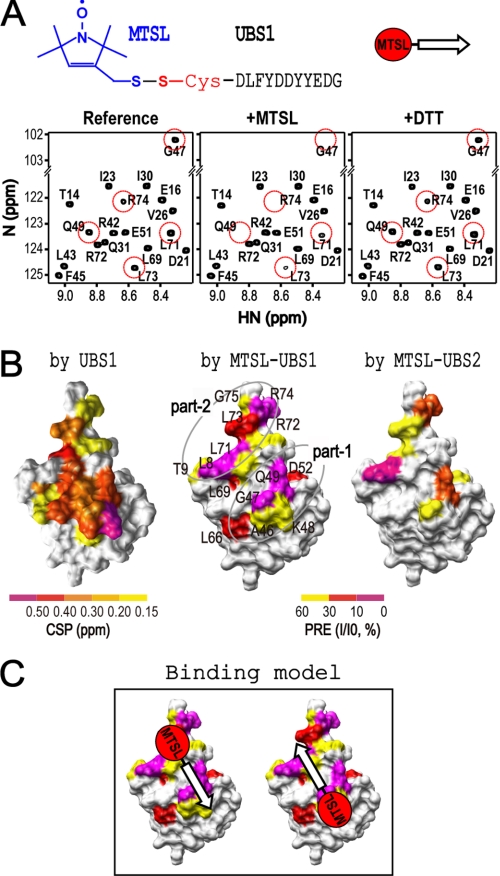
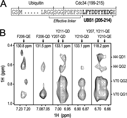
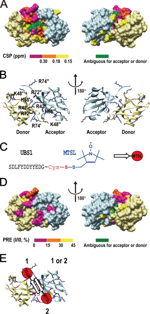
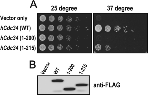
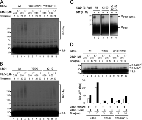
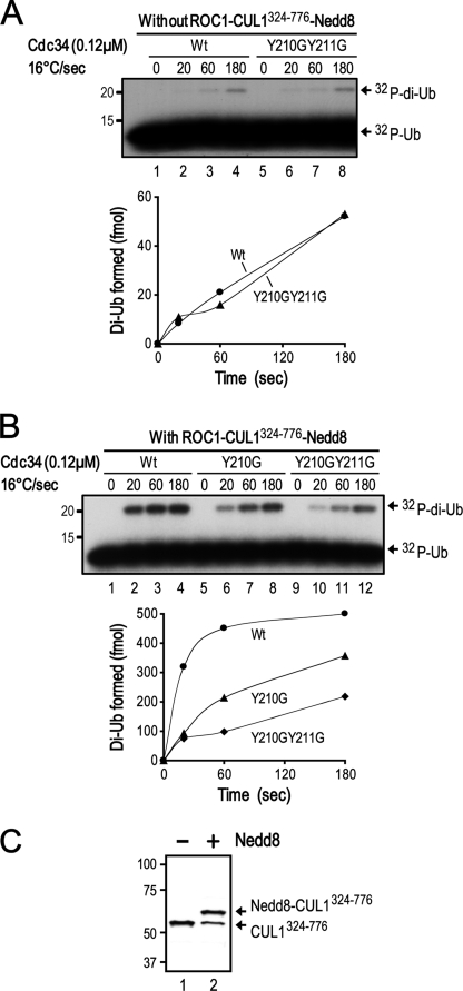
Cdc34 is an E2 ubiquitin-conjugating enzyme that functions in conjunction with SCF (Skp1.Cullin 1.F-box) E3 ubiquitin ligase to catalyze covalent attachment of polyubiquitin chains to a target protein. Here we identified direct interactions between the human Cdc34 C terminus and ubiquitin using NMR chemical shift perturbation assays. The ubiquitin binding activity was mapped to two separate Cdc34 C-terminal motifs (UBS1 and UBS2) that comprise residues 206-215 and 216-225, respectively. UBS1 and UBS2 bind to ubiquitin in the proximity of ubiquitin Lys(48) and C-terminal tail, both of which are key sites for conjugation. When bound to ubiquitin in one orientation, the Cdc34 UBS1 aromatic residues (Phe(206), Tyr(207), Tyr(210), and Tyr(211)) are probably positioned in the vicinity of ubiquitin C-terminal residue Val(70). Replacement of UBS1 aromatic residues by glycine or of ubiquitin Val(70) by alanine decreased UBS1-ubiquitin affinity interactions. UBS1 appeared to support the function of Cdc34 in vivo because human Cdc34(1-215) but not Cdc34(1-200) was able to complement the growth defect by yeast Cdc34 mutant strain. Finally, reconstituted IkappaBalpha ubiquitination analysis revealed a role for each adjacent pair of UBS1 aromatic residues (Phe(206)/Tyr(207), Tyr(210)/Tyr(211)) in conjugation, with Tyr(210) exhibiting the most pronounced catalytic function. Intriguingly, Cdc34 Tyr(210) was required for the transfer of the donor ubiquitin to a receptor lysine on either IkappaBalpha or a ubiquitin in a manner that depended on the neddylated RING sub-complex of the SCF. Taken together, our results identified a new ubiquitin binding activity within the human Cdc34 C terminus that contributes to SCF-dependent ubiquitination.


Figures

FIGURE 1.
FIGURE 1.
Identification of the hCdc34C UBS1 and UBS2 motifs for binding to ubiquitin. A, the CSP experiments were carried out with 15N-labeled hCdc34C (0.1 mm) in the presence of ubiquitin (1.5 mm) in a buffer containing 50 mm MES (pH 5.5) or Tris-HCl (pH 7.5), 100 mm NaCl, and 5 mm DTT. The bar graphs show the CSP signals (at pH 5.5 or 7.5) measured across the entire hCdc34C region, exhibiting two regions designated as UBS1 and UBS2. The Kd values of UBS1 and UBS2 are indicated. B, the plots show the averaged CSP signal determined for 15N-labeled hCdc34C (0.1 mm) in the presence of ubiquitin (1.5 mm) at a range of pH (left, with 100 mm NaCl) or salt concentration (right, at pH 5.5). C, the CSP experiments were carried out with 15N-labeled ubiquitin (0.2 mm) in the presence of increasing concentrations of hCdc34C in a buffer containing 50 mm MES (pH 5.5), 100 mm NaCl, and 5 mm DTT. The plot shows the CSP response at various ubiquitin residues (as specified) as a function of hCdc34C concentration. D, the hCdc34C-binding surface on 15N-labeled ubiquitin is shown, with different colors denoting the CSP intensity. E, sequence alignment of the C-terminal regions of Cdc34 homologs. The red, blue, and green colors represent residues that are identical, strongly conserved, and conserved, respectively. Residues 187–196 and the UBS1 and UBS2 regions in Cdc34C are highlighted in yellow, cyan, and gray, respectively.
FIGURE 2.
FIGURE 2.
PRE analysis of the binding surface and orientation of UBS1 or UBS2 on ubiquitin. All NMR experiments were performed in a buffer containing 50 mm MES (pH 5.5) and 100 mm NaCl. A, top, schematic presentation of the MTLS-labeled UBS1 peptide. Note that this peptide contains an N-terminal Cys residue for spin labeling. Bottom, the HSQC spectra of 15N-labeled ubiquitin (0.1 mm) were recorded in the absence (Reference) and presence (+MTSL) of the MTSL-UBS1 peptide, respectively. In a separate sample (+DTT), DTT (10 mm) was incubated with 15N-labeled ubiquitin and MTSL-UBS1 overnight. The detailed procedure is described under “Experimental Procedures.” B, left, the CSP surface of 15N-labeled ubiquitin (0.2 mm) in the presence of the UBS1 peptide (1.5 mm) was determined and is shown based on CSP intensity. Middle, the paramagnetic relaxation enhanced residues on 15N-labeled ubiquitin in the presence of the MTSL-UBS1 peptide are shown. The paramagnetic relaxation-enhanced regions can be divided into two separated regions, in which “part 1” contains Ala46, Gly47, and Gln49, and “part 2” contains Leu8, Leu71, Leu73, and Arg74. Right, the same PRE experiment was performed for the MSTL-UBS2 peptide. UBS2 contains Cys (GEVEEEADSCFG), which can react with MTSL. The paramagnetic relaxation-enhanced regions by the MTSL-UBS2 peptide are very similar to those enhanced by the MTSL-UBS1 peptide. C, models for the dual ubiquitin binding orientations for MTSL-UBS1.
FIGURE 3.
FIGURE 3.
NOE cross-peak analysis of the interactions between UBS1 and ubiquitin. A, a diagram for the ubiquitin linker-UBS1 fusion protein. B, NOE cross-peaks were observed in 13C-resolved three-dimensional aromatic NOESY-HSQC spectrum of 13C/15N-labeled ubiquitin linker-UBS1 hybrid protein. Ubiquitin Val70 and Ile44 had NOE cross-peaks with all of the UBS1 aromatic residues (Phe206, Tyr207, Tyr210, and Tyr211).
FIGURE 4.
FIGURE 4.
UBS1 binds to Lys48-diubiquitin. The NMR experiments were carried out in a buffer containing 50 mm MES (pH 5.5) and 100 mm NaCl. A, CSP analysis. The UBS1-binding surface on 15N-labeled Lys48-linked diubiquitin is shown, with different colors denoting the CSP intensity. B, ribbon representations of Lys48- diubiquitin. The basic amino acids in the diubiquitin interface, such as Lys, Arg, and His, are differently labeled according to their originations. Single and double quotation marks represent the residues from acceptor and donor ubiquitin, respectively. C and D, PRE analysis. C, schematic representations of the UBS1-MTSL peptide. D, PRE experiments were carried out with 15N-labeled diubiquitin (0.1 mm) and UBS1-MTSL. Shown is the UBS1-MTSL-induced enhancement of the relaxation regions in diubiquitin. E, a model shows the dual binding orientation of the UBS1-MTSL peptide on Lys48-diubiquitin.
FIGURE 5.
FIGURE 5.
The UBS1-containing hCdc34(1–215) rescues cell viability in yeast. A, growth assay. Colony growth is shown for the temperature-sensitive cdc34-2 yeast strain transformed with vectors expressing the wild type hCdc34 or truncated forms, as indicated, at permissive (left) or non-permissive (right) temperature. The experimental details are described under “Experimental Procedures.” B, immunoblot analysis. Galactose-induced whole yeast cell extracts were prepared and analyzed by anti-FLAG Western blot to monitor the expression of hCdc34, hCdc34(1–200), and hCdc34(1–215).
FIGURE 6.
FIGURE 6.
The hCdc34 UBS1 aromatic residues contribute to SCF-dependent and Cdc34-catalyzed ubiquitination. The polyubiquitination (A and B) or monoubiquitination (D) of IκBα was carried out in the presence of the wild type or mutant form of hCdc34 as indicated. The reaction was incubated for the times specified. The influence of the hCdc34 mutation on the production of the monoubiquitinated species was quantified and is presented graphically. UbK0, lysineless ubiquitin. C, E2-S∼ubiquitin formation assay was carried out as described under “Experimental Procedures.”
FIGURE 7.
FIGURE 7.
The UBS1 Y210G/Y211G mutation is defective in diubiquitin synthesis that requires the neddylated RING subcomplex of SCF. The diubiquitin synthesis reaction was carried out in the absence (A) or presence (B) of the neddylated ROC1-CUL1(324–776). The influence of the hCdc34 mutation on the production of diubiquitin was quantified and is presented graphically. C, immunoblot analysis of an aliquot of the neddylation reaction mixture (as described under “Experimental Procedures”).

Similar articles

Cited by

References

    1. Hershko A., Ciechanover A. (1998) Annu. Rev. Biochem. 67, 425–479 - PubMed
    1. Pickart C. M. (2004) Cell 116, 181–190 - PubMed
    1. Feldman R. M., Correll C. C., Kaplan K. B., Deshaies R. J. (1997) Cell 91, 221–230 - PubMed
    1. Skowyra D., Craig K. L., Tyers M., Elledge S. J., Harper J. W. (1997) Cell 91, 209–219 - PubMed
    1. Tan P., Fuchs S. Y., Chen A., Wu K., Gomez C., Ronai Z., Pan Z. Q. (1999) Mol. Cell 3, 527–533 - PubMed

Publication types