. 2010 May 28;285(22):16830-43.
doi: 10.1074/jbc.M110.106187. Epub 2010 Mar 29.

AP-1 (Fra-1/c-Jun)-mediated induction of expression of matrix metalloproteinase-2 is required for 15S-hydroxyeicosatetraenoic acid-induced angiogenesis

Affiliations

AP-1 (Fra-1/c-Jun)-mediated induction of expression of matrix metalloproteinase-2 is required for 15S-hydroxyeicosatetraenoic acid-induced angiogenesis

Nikhlesh K Singh et al. J Biol Chem. .

Abstract

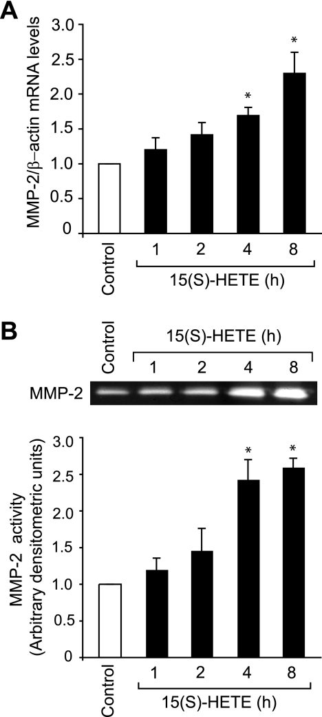
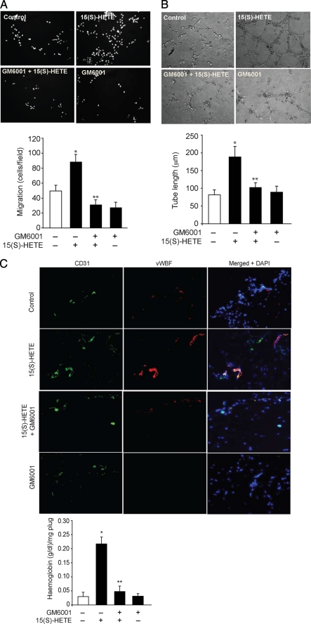
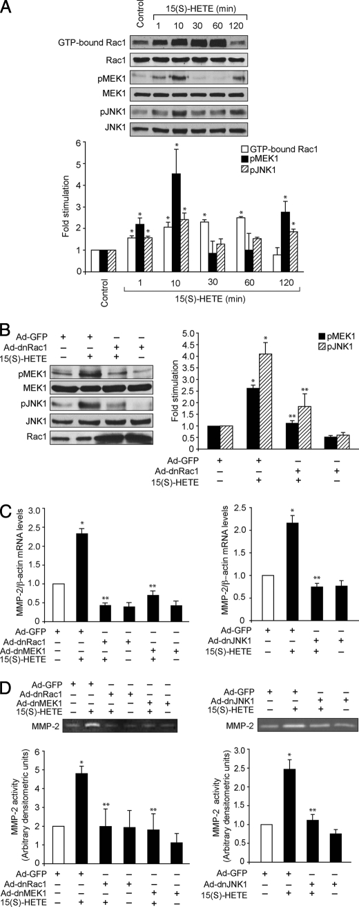
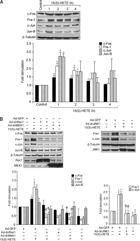
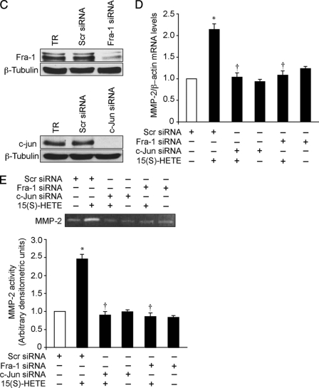
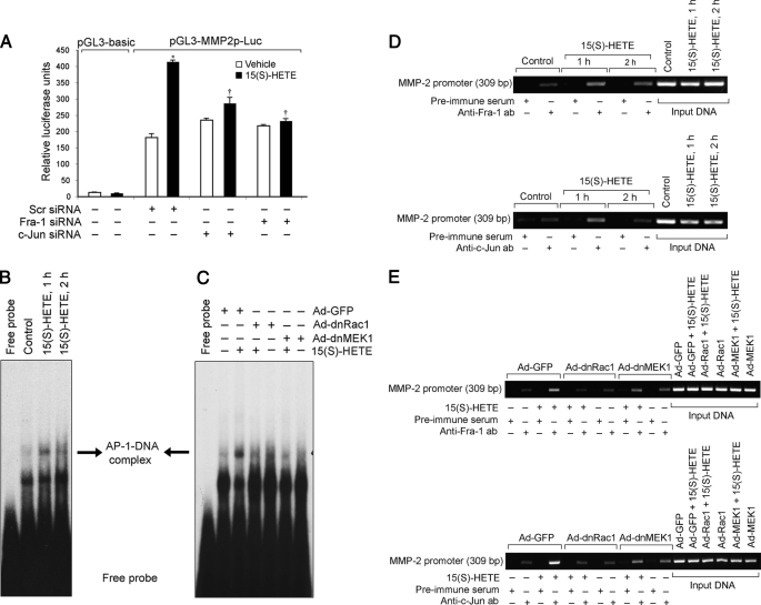
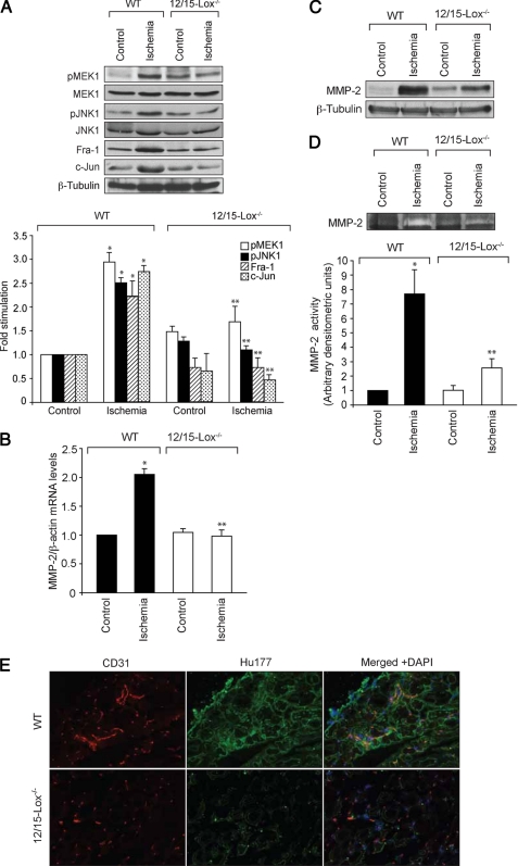
To understand the involvement of matrix metalloproteinases (MMPs) in 15(S)-hydroxyeicosatetraenoic acid (15(S)-HETE)-induced angiogenesis, we have studied the role of MMP-2. 15(S)-HETE induced MMP-2 expression and activity in a time-dependent manner in human dermal microvascular endothelial cells (HDMVECs). Inhibition of MMP-2 activity or depletion of its levels attenuated 15(S)-HETE-induced HDMVEC migration, tube formation, and Matrigel plug angiogenesis. 15(S)-HETE also induced Fra-1 and c-Jun expression in a Rac1-MEK1-JNK1-dependent manner. In addition, 15(S)-HETE-induced MMP-2 expression and activity were mediated by Rac1-MEK1-JNK1-dependent activation of AP-1 (Fra-1/c-Jun). Cloning and site-directed mutagenesis of MMP-2 promoter revealed that AP-1 site proximal to the transcriptional start site is required for 15(S)-HETE-induced MMP-2 expression, and Fra-1 and c-Jun are the essential components of AP-1 that bind to MMP-2 promoter in response to 15(S)-HETE. Hind limb ischemia led to an increase in MEK1 and JNK1 activation and Fra-1, c-Jun, and MMP-2 expression resulting in enhanced neovascularization and recovery of blood perfusion in wild-type mice as compared with 12/15-Lox(-/-) mice. Together, these results provide the first direct evidence for a role of 12/15-Lox-12/15(S)-HETE axis in the regulation of ischemia-induced angiogenesis.


Figures

FIGURE 1.
FIGURE 1.
15(S)-HETE induces MMP-2 expression and activity. A and B, quiescent HDMVECs were treated with and without 0.1 μm 15(S)-HETE for the indicated time periods, and either total cellular RNA was isolated and analyzed for MMP-2 and β-actin mRNA levels by QRT-PCR (A) or medium was collected and analyzed for MMP-2 activity by gelatin zymography (B). The bar graphs in A and B represent the mean ± S.D. values of three independent experiments. *, p < 0.01 versus control.
FIGURE 2.
FIGURE 2.
MMP-2 mediates 15(S)-HETE-induced HDMVEC migration and tube formation in vitro and Matrigel plug angiogenesis in vivo. A and B, quiescent HDMVECs were treated with and without 10 mm GM6001 for 30 min at 37 °C, trypsinized, rinsed with trypsin-neutralizing solution, and subjected to 0.1 μm 15(S)-HETE-induced migration (A) or tube formation (B). C, WT mice were injected subcutaneously with 0.5 ml of Matrigel premixed with vehicle or 10 μm 15(S)-HETE with and without 10 mm GM6001. One week later, the animals were sacrificed, and the Matrigel plugs were harvested from underneath the skin, and either cryosections were made and examined by double immunofluorescence staining for vWF and CD31 using their specific antibodies or analyzed for hemoglobin content using Drabkin's reagent. D, HDMVECs were transfected with scrambled or MMP-2 siRNA, and 48 h later cell extracts were prepared and analyzed by Western blotting for MMP-2 levels using its specific antibodies. E and F, all the conditions were the same as in D except that, after transfection, HDMVECs were quiesced and subjected to 15(S)-HETE (0.1 μm)-induced migration (E) or tube formation (F). The bar graphs in A–F represent the mean ± S.D. values of three independent experiments or six plugs from six animals. *, p < 0.01 versus control; **, p < 0.01 versus 15(S)-HETE. TR, transfection reagent.
FIGURE 2.
FIGURE 2.
MMP-2 mediates 15(S)-HETE-induced HDMVEC migration and tube formation in vitro and Matrigel plug angiogenesis in vivo. A and B, quiescent HDMVECs were treated with and without 10 mm GM6001 for 30 min at 37 °C, trypsinized, rinsed with trypsin-neutralizing solution, and subjected to 0.1 μm 15(S)-HETE-induced migration (A) or tube formation (B). C, WT mice were injected subcutaneously with 0.5 ml of Matrigel premixed with vehicle or 10 μm 15(S)-HETE with and without 10 mm GM6001. One week later, the animals were sacrificed, and the Matrigel plugs were harvested from underneath the skin, and either cryosections were made and examined by double immunofluorescence staining for vWF and CD31 using their specific antibodies or analyzed for hemoglobin content using Drabkin's reagent. D, HDMVECs were transfected with scrambled or MMP-2 siRNA, and 48 h later cell extracts were prepared and analyzed by Western blotting for MMP-2 levels using its specific antibodies. E and F, all the conditions were the same as in D except that, after transfection, HDMVECs were quiesced and subjected to 15(S)-HETE (0.1 μm)-induced migration (E) or tube formation (F). The bar graphs in A–F represent the mean ± S.D. values of three independent experiments or six plugs from six animals. *, p < 0.01 versus control; **, p < 0.01 versus 15(S)-HETE. TR, transfection reagent.
FIGURE 3.
FIGURE 3.
Rac1, MEK1, and JNK1 mediate 15(S)-HETE-induced MMP-2 expression and activity. A, quiescent HDMVECs were treated with and without 0.1 μm 15(S)-HETE for the indicated time periods, and cell extracts were prepared and analyzed for Rac1 activation by pulldown assay and MEK1 and JNK1 phosphorylation by Western blotting using their phosphospecific antibodies. Whereas total cellular levels of Rac1 are shown in the second blot from the top, the pMEK1 and pJNK1 blots were reprobed with anti-MEK1 and anti-JNK1 antibodies for normalization. B, HDMVECs were transduced with Ad-GFP or Ad-dnRac1 at 40 m.o.i., quiesced, treated with and without 15(S)-HETE (0.1 μm) for 10 min, and MEK1 and JNK1 phosphorylation were measured. The blots were reprobed with anti-MEK1 or anti-JNK1 antibodies for normalization. One of these blots was reprobed with anti-Rac1 antibodies to show the overexpression of dnRac1. C and D, HDMVECs were transduced with Ad-GFP, Ad-dnRac1, Ad-dnMEK1, or Ad-dnJNK1 at 40 m.o.i., quiesced, treated with and without 15(S)-HETE (0.1 μm) for 8 h and either total cellular RNA was isolated and analyzed for MMP-2 and β-actin mRNA levels by QRT-PCR (C) or medium was collected and assayed for MMP-2 activity by gelatin zymography (D). The bar graphs in A–D represent mean ± S.D. values of three independent experiments. *, p < 0.01 versus control or Ad-GFP; **, p < 0.01 versus 15(S)-HETE or Ad-GFP plus 15(S)-HETE.
FIGURE 4.
FIGURE 4.
AP-1 (Fra-1/c-Jun) mediates 15(S)-HETE-induced MMP-2 expression and activity. A, quiescent HDMVECs were treated with and without 15(S)-HETE (0.1 μm) for the indicated time periods, and cell extracts were prepared and analyzed by Western blotting for c-Fos, Fra-1, c-Jun, Jun-B, and β-tubulin levels using their respective antibodies. B, HDMVECs were transduced with Ad-GFP, Ad-dnRac1, Ad-dnMEK1, or Ad-dnJNK1 at 40 m.o.i., quiesced, treated with and without 15(S)-HETE (0.1 μm) for 1 h, and cell extracts were prepared and analyzed for either c-Fos, Fra-1, c-Jun, Jun-B, and β-tubulin levels or Fra-1 and c-Jun levels as described in A. The c-Jun blot was reprobed sequentially with anti-Rac1, anti-MEK1, or anti-JNK1 antibodies to show the overexpression of dnRac1, dnMEK1, and dnJNK1, respectively. C, HDMVECs were transfected with scrambled, Fra-1, or c-Jun siRNA, and 48 h later cell extracts were prepared and analyzed by Western blotting for Fra-1 and c-Jun levels using their specific antibodies. D and E, HDMVECs were transfected with scrambled, Fra-1, or c-Jun siRNA, quiesced, treated with and without 15(S)-HETE (0.1 μm) for 8 h, and either total cellular RNA was isolated and analyzed for MMP-2 and β-actin mRNA levels by QRT-PCR (D) or medium was collected and analyzed for MMP-2 activity by gelatin zymography (E). The bar graphs in A–E represent mean ± S.D. values of three independent experiments. *, p < 0.01 versus control or Ad-GFP or scrambled siRNA; , p < 0.01 versus 15(S)-HETE or Ad-GFP plus 15(S)-HETE or scrambled siRNA plus 15(S)-HETE.
FIGURE 4.
FIGURE 4.
AP-1 (Fra-1/c-Jun) mediates 15(S)-HETE-induced MMP-2 expression and activity. A, quiescent HDMVECs were treated with and without 15(S)-HETE (0.1 μm) for the indicated time periods, and cell extracts were prepared and analyzed by Western blotting for c-Fos, Fra-1, c-Jun, Jun-B, and β-tubulin levels using their respective antibodies. B, HDMVECs were transduced with Ad-GFP, Ad-dnRac1, Ad-dnMEK1, or Ad-dnJNK1 at 40 m.o.i., quiesced, treated with and without 15(S)-HETE (0.1 μm) for 1 h, and cell extracts were prepared and analyzed for either c-Fos, Fra-1, c-Jun, Jun-B, and β-tubulin levels or Fra-1 and c-Jun levels as described in A. The c-Jun blot was reprobed sequentially with anti-Rac1, anti-MEK1, or anti-JNK1 antibodies to show the overexpression of dnRac1, dnMEK1, and dnJNK1, respectively. C, HDMVECs were transfected with scrambled, Fra-1, or c-Jun siRNA, and 48 h later cell extracts were prepared and analyzed by Western blotting for Fra-1 and c-Jun levels using their specific antibodies. D and E, HDMVECs were transfected with scrambled, Fra-1, or c-Jun siRNA, quiesced, treated with and without 15(S)-HETE (0.1 μm) for 8 h, and either total cellular RNA was isolated and analyzed for MMP-2 and β-actin mRNA levels by QRT-PCR (D) or medium was collected and analyzed for MMP-2 activity by gelatin zymography (E). The bar graphs in A–E represent mean ± S.D. values of three independent experiments. *, p < 0.01 versus control or Ad-GFP or scrambled siRNA; , p < 0.01 versus 15(S)-HETE or Ad-GFP plus 15(S)-HETE or scrambled siRNA plus 15(S)-HETE.
FIGURE 5.
FIGURE 5.
15(S)-HETE-induced MMP-2 promoter-luciferase reporter gene activity requires −1263 AP-1-binding element. A, sequence of the cloned 1.7-kb human MMP-2 promoter showing the −1263, −1615, and −1673 AP-1-binding sites. B, HDMVECs were transfected with empty vector (pGL3-basic) or MMP-2 promoter-luciferase constructs with and without site-directed mutagenesis of AP-1 sites, quiesced, and treated with and without 15(S)-HETE for 8 h, and cell extracts were prepared and analyzed for luciferase activity. M1, M2, and M3 indicate mutations in AP-1 binding sites located at −1263, −1615, and −1673 nt, respectively. *, p < 0.01 versus vehicle control; , p < 0.01 versus pGL3-MMP2p-Luc plus 15(S)-HETE. Dotted vertical lines indicate mutated AP-1 sites.
FIGURE 6.
FIGURE 6.
Fra-1 and c-Jun bind to MMP-2 promoter in response to 15(S)-HETE in a Rac1- and MEK1-dependent manner. A, HDMVECs were transfected with scrambled, Fra-1, or c-Jun siRNA in combination with empty vector or pGL3-MMP2p-Luc, quiesced, treated with and without 0.1 μm 15(S)-HETE for 8 h, and cell extracts were prepared and analyzed for luciferase activity. B and D, quiescent HDMVECs were treated with and without 15(S)-HETE (0.1 μm) for the indicated time periods, and either nuclear extracts were prepared and analyzed by EMSA for AP-1-binding activity using 32P-labeled −1263 AP-1 binding sequence of MMP-2 promoter as a probe in vitro (B) or processed for ChIP analysis of Fra-1 and c-Jun binding to MMP-2 promoter in vivo using primers encompassing −1263 AP-1 site (D). C and E, HDMVECs were transduced with Ad-GFP, Ad-dnRac1, or Ad-dnMEK1 at 40 m.o.i., quiesced, and treated with and without 15(S)-HETE (0.1 μm) for 1 h, and either nuclear extracts were prepared and analyzed by EMSA for −1263 AP-1-binding activity as described in B (C) or processed for ChIP analysis of Fra-1 and c-Jun binding to MMP-2 promoter in vivo as described in D (E). *, p < 0.01 versus vehicle control; , p < 0.01 versus pGL3-MMP2p-Luc plus 15(S)-HETE.
FIGURE 7.
FIGURE 7.
Lack of 12/15-Lox gene impairs blood flow recovery after ischemia. WT and 12/15-Lox−/− mice were subjected to hind limb ischemia by left femoral artery excision. A, on day 7 after hind limb ischemia, blood flow was measured by using laser Doppler perfusion imaging. Perfusion is expressed as the ratio of the ischemic to the non-ischemic hind limb. B, blood vessels in the ischemic adductor muscles of WT and 12/15-Lox−/− mice were analyzed by double immunofluorescence staining for CD31 (green) and vWF (red). *, p < 0.01 versus WT mice.
FIGURE 8.
FIGURE 8.
Lack of 12/15-Lox gene impairs ischemia-induced MMP-2 expression and activity. A–D, adductor muscles were isolated from ischemic and non-ischemic WT and 12/15-Lox−/− mice 7 days post-operation, and either tissue extracts were prepared or RNA was isolated. Tissue extracts were analyzed by Western blotting for MEK1 and JNK1 phosphorylation and Fra-1 and c-Jun levels using the respective antibodies (A). MMP-2 mRNA levels were measured by QRT-PCR (B), and its protein levels and activity were measured by Western blotting and gelatin zymography, respectively (C and D). E, cryptic collagen epitopes were evaluated in the ischemic adductor muscles of WT and 12/15-Lox−/− mice by immunofluorescence staining with anti-Hu177 antibodies (green) and anti-CD31 antibodies (red). The bar graphs in A, B, and D represent the mean ± S.D. values for six animals. *, p < 0.01 versus WT mice.

Similar articles

Cited by

References

    1. Folkman J. (1995) Nat. Med. 1, 27–31 - PubMed
    1. Freedman S. B., Isner J. M. (2001) J. Mol. Cell Cardiol. 33, 379–393 - PubMed
    1. Mignatti P., Rifkin D. B. (1996) Enzyme Protein 49, 117–137 - PubMed
    1. Lauer-Fields J. L., Sritharan T., Stack M. S., Nagase H., Fields G. B. (2003) J. Biol. Chem. 278, 18140–18145 - PubMed
    1. Gross J., Lapiere C. M. (1962) Proc. Natl. Acad. Sci. U.S.A. 48, 1014–1022 - PMC - PubMed

Publication types

Substances