Comparative Study
. 2010 Apr;133(Pt 4):1054-66.
doi: 10.1093/brain/awq028. Epub 2010 Mar 30.

T cells can mediate viral clearance from ependyma but not from brain parenchyma in a major histocompatibility class I- and perforin-independent manner

Affiliations
Comparative Study

T cells can mediate viral clearance from ependyma but not from brain parenchyma in a major histocompatibility class I- and perforin-independent manner

Daniel D Pinschewer et al. Brain. 2010 Apr.

Abstract

Viral infection of the central nervous system can lead to disability and death. Yet the majority of viral infections with central nervous system involvement resolve with only mild clinical manifestations, if any. This is generally attributed to efficient elimination of the infection from the brain coverings, i.e. the meninges, ependyma and chorioplexus, which are the primary targets of haematogeneous viral spread. How the immune system is able to purge these structures from viral infection with only minimal detrimental effects is still poorly understood. In the present work we studied how an attenuated lymphocytic choriomeningitis virus can be cleared from the central nervous system in the absence of overt disease. We show that elimination of the virus from brain ependyma, but not from brain parenchyma, could be achieved by a T cell-dependent mechanism operating independently of major histocompatibility class I antigens and perforin. Considering that cytotoxic T lymphocyte-mediated cytotoxicity is a leading cause of viral immunopathology and tissue damage, our findings may explain why the most common viral intruders of the central nervous system rarely represent a serious threat to our health.


Figures
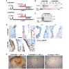
Figure 1
Figure 1
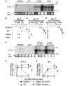
Genome organization, viral spread and blood brain barrier integrity following intracerebral infection with either LCMV Armstrong or rLCMV/INDG. (A) Schema of the LCMV-Armstrong (LMCV-ARM) and rLCMV/INDG genomes. Both viruses consist of two single-stranded negative-strand RNA segments, encoding two viral genes in ambisense orientation each. The long (L) segment encodes for the RNA-dependent RNA polymerase L and for the matrix protein Z, while the short (S) segment carries the GP and NP genes. rLCMV/INDG was created by substituting the LCMV-GP gene for vesicular stomatitis virus-INDG (Pinschewer et al., 2003). (B–E) C57BL/6 mice were infected intracerebrally with rLCMV/INDG or LCMV-ARM as indicated or were left uninfected. (B and C) Viral S-segment (S seg.) RNA in brain was detected at the indicated day (d) after intracerebral infection by northern blot (ethidium bromide staining of 28S rRNA indicates loading control; lanes represent individual animals). (D) Immunohistochemical detection of LCMV-NP confirms reduced spread of rLCMV/INDG as compared to LCMV-Armstrong. Note that LCMV- Armstrong (Days 4 and 6) and also rLCMV/INDG (Day 6) spreads into some subependymal cells (arrows). (E) At Day 6 after infection, animals were given horseradish peroxide (HRP) intravenously. One hour later, they were sacrificed for detection of horseradish peroxidase leakage into the brain parenchyma, indicative of blood brain barrier breakdown. Representative pictures of horseradish peroxidase reactions are shown (n = 3–4 animals per group). Scale bar for D: 100 µm; for E: 500 µm.
Figure 2
Figure 2
Absence of disease after rLCMV/INDG intracerebral infection is not due to altered antiviral cytotoxic T cell response. (A) C57BL/6 mice were infected by the intravenous and intracerebral route with LCMV-Armstrong (LCMV-ARM) and/or rLCMV/INDG in various combinations as indicated in the chart, summarizing also the clinical outcome of infection. Animals exhibiting clinical signs of terminal choriomeningitis were euthanized in accordance with the Swiss law for animal protection. (B) NP396-specific (expressed by both viruses) and GP33-specific (expressed by LCMV-ARM) CD8+ T cells in blood were enumerated on Day 6 by MHC class I tetramer staining. (C) Primary ex vivo cytotoxic T cell activity of splenocytes against NP396 was tested 6 days after infection. Symbols represent the mean ± SEM of three mice per group. Symbol keys are provided in the table of panel A. i.c. = intracerebral; i.v. = intravenous.
Figure 3
Figure 3
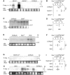
T cells—but not antibodies—are necessary for clearance of rLCMV/INDG from the CNS. Mice of the indicated genotypes were infected with rLCMV/INDG intracerebrally. (A) Detection of viral S-segment (S seg.) RNA in the brain by northern blot on Day 14 (ethidium bromide staining of 28S rRNA indicates loading control). (B) Frequencies of NP396-specific CD8+ T cells in blood were determined on Day 8 using MHC class I tetramers. TCRβδ−/− and RAG−/− mice lack CD8+ T cells. (C and D) Virus-neutralizing total Ig (C) and IgG (D) were determined at the indicated time points. (E–G) C57BL/6, JHT and TCRβδ−/− mice were infected as above. An additional group of TCRβδ−/− mice was treated with 500 µl of vesicular stomatitis virus-immune serum on Day 7. (E) Viral RNA in the brain was detected on Day 14 by northern hybridization. (F and G) Virus neutralizing total Ig and IgG titres in serum were determined over time. Lanes in A and E and symbols in B–D represent individual mice. Symbols in F–G represent the mean ± SEM of three mice per group. Representative results from two similar experiments are shown. n.d. = not detectable; n.s. = not significant (P > 0.05); ** = highly significant (P < 0.01).
Figure 4
Figure 4
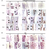
Clearance of LCMV/INDG from the CNS is dependent on MHC class I and perforin. Mice of the indicated genotypes were infected with rLCMV/INDG intracerebrally. (A, C, E, G and H) Viral S-segment (S seg.) RNA was detected by northern blot on Day 14 after intracerebral infection. Ethidium bromide staining is shown as loading control. (B, D, F and I) NP396-specific CD8+ T cell frequencies in blood were enumerated at Day 7 after infection. MHCI−/− mice and CD8−/− mice lack CD8+ T cells. Symbols and lanes represent individual mice. In figure F and I, multiple comparisons were not performed since the F-test of ANOVA failed to detect significant differences (P > 0.05). n.d. = not detectable; n.s. = not significant (P > 0.05).
Figure 5
Figure 5
rLCMV/INDG dissemination in the brain of perforin-deficient, TCRβδ−/− and RAG−/− mice, and dense CD8+ T cell infiltrates in perforin-deficient mice. Perforin-deficient (PKO), TCRβδ−/−, RAG−/− and C57BL/6 wild-type control mice were infected with rLCMV/INDG intracerebrally and were sacrificed at the indicated time points. Brain tissues were processed for immunohistochemical analysis of LCMV NP (A) and CD8+ T cells (B). The latter analysis was only performed for perforin-deficient and C57BL/6 mice since TCRβδ−/- and RAG−/− mice lack T cells. Representative images of 2–4 animals per group and timepoint are shown. Arrowheads in (A) indicate infected (LCMV NP-positive) subependymal cells. Insets in the Day 12 timepoints of (A) show persisting infection of ependymal cells in TCRβδ−/− and RAG−/− but not in perforin-deficient mice, whereas subependymal cells are infected in all three knockout strains. Scale bars A: 100 µm; B: 50 µm. d = days post intracerebral infection.
Figure 6
Figure 6
The cell type-specific distribution of rLCMV/INDG in the CNS is different in TCRβδ−/− and RAG−/− mice as compared to perforin-deficient and MHCI−/− mice. Mice of the indicated genotypes were infected with rLCMV/INDG intracerebrally. Fourteen days later they were sacrificed and brain tissues were processed for histological analysis of LCMV NP. (A) Ependyma was differentiated based on morphological criteria (arrows in top panel) combined with immunohistochemical detection of LCMV NP (brown). Infection of parenychmal cell types (bottom panels) was detected by combining LCMV NP staining (red) with cell type-specific markers (green) in immunofluorescent co-staining as indicated. Arrowheads point out colocalization of cell type-specific markers with LCMV NP in immunofluorescence images. Representative images from three to four animals per group are shown. Scale bar: 50 µm. (B) Histological images were quantified to define the proportion of each cell type within the total population of infected cells. (C) Viral RNA loads in the brain of the indicated mouse strains were measured by quantitative RT-PCR and are displayed in arbitrary units (see ‘Materials and methods’ section). Bars represent the mean + SD of four to seven animals per group. Viral RNA loads in infected C57BL/6 mice were significantly different from perforin-deficient (PKO), MHCI−/−, TCRβδ−/− and RAG−/− mice (P < 0.01), whereas the latter four were not significantly different from each other (P > 0.05 for all comparisons). The technical background was determined in brain tissue of uninfected mice (mean of six animals indicated as dashed line), and was not significantly different from infected C57BL/6 wild-type mice (P > 0.05, not shown).

Similar articles

Cited by

References

    1. Adachi M, Suematsu S, Kondo T, Ogasawara J, Tanaka T, Yoshida N, et al. Targeted mutation in the Fas gene causes hyperplasia in peripheral lymphoid organs and liver. Nat Genet. 1995;11:294–300. - PubMed
    1. Adair CV, Gauld RL, Smadel JE. Aseptic meningitis, a disease of diverse etiology: clinical and etiologic studies on 854 cases. Ann Intern Med. 1953;39:675–704. - PubMed
    1. Ahmed R, Gray D. Immunological memory and protective immunity: understanding their relation. Science. 1996;272:54–60. - PubMed
    1. Andersen IH, Marker O, Thomsen AR. Breakdown of blood-brain barrier function in the murine lymphocytic choriomeningitis virus infection mediated by virus-specific CD8+ T cells. J Neuroimmunol. 1991;31:155–63. - PubMed
    1. Badovinac VP, Porter BB, Harty JT. Programmed contraction of CD8(+) T cells after infection. Nat Immunol. 2002;3:619–26. - PubMed

Publication types

MeSH terms

Substances