. 2010 Jun;42(1):23-41.
doi: 10.1152/physiolgenomics.00027.2010. Epub 2010 Mar 30.

Identifying physiological origins of baroreflex dysfunction in salt-sensitive hypertension in the Dahl SS rat

Affiliations

Identifying physiological origins of baroreflex dysfunction in salt-sensitive hypertension in the Dahl SS rat

Scott M Bugenhagen et al. Physiol Genomics. 2010 Jun.

Abstract

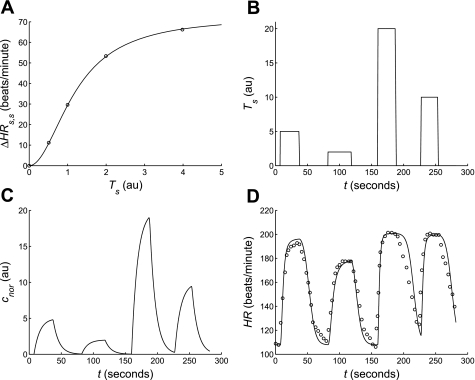
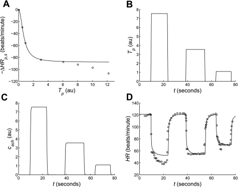
Salt-sensitive hypertension is known to be associated with dysfunction of the baroreflex control system in the Dahl salt-sensitive (SS) rat. However, neither the physiological mechanisms nor the genomic regions underlying the baroreflex dysfunction seen in this rat model are definitively known. Here, we have adopted a mathematical modeling approach to investigate the physiological and genetic origins of baroreflex dysfunction in the Dahl SS rat. We have developed a computational model of the overall baroreflex heart rate control system based on known physiological mechanisms to analyze telemetry-based blood pressure and heart rate data from two genetic strains of rat, the SS and consomic SS.13(BN), on low- and high-salt diets. With this approach, physiological parameters are estimated, unmeasured physiological variables related to the baroreflex control system are predicted, and differences in these quantities between the two strains of rat on low- and high-salt diets are detected. Specific findings include: a significant selective impairment in sympathetic gain with high-salt diet in SS rats and a protection from this impairment in SS.13(BN) rats, elevated sympathetic and parasympathetic offsets with high-salt diet in both strains, and an elevated sympathetic tone with high-salt diet in SS but not SS.13(BN) rats. In conclusion, we have associated several important physiological parameters of the baroreflex control system with chromosome 13 and have begun to identify possible physiological mechanisms underlying baroreflex impairment and hypertension in the Dahl SS rat that may be further explored in future experimental and modeling-based investigation.


Figures

Fig. 1.
Fig. 1.
Overall baroreflex flow diagram. The various subcomponents comprising the model of the overall baroreflex control of heart rate are shown. Aortic blood pressure serves as the model input and is transduced to a neural signal (firing rate) by afferent baroreceptors found in the aortic arch. This neural signal is further processed by the central nervous system (CNS). The output of the CNS serves to modulate the activities (outflows) of the 2 parallel pathways of the peripheral nervous system (sympathetic and parasympathetic). These 2 pathways exert reciprocal effects at the sinoatrial (SA) node of the heart, which is the ultimate effector of heart rate. Heart rate is the final output of the model.
Fig. 2.
Fig. 2.
Pressure-radius relationship of rat aorta. The radius (R) of the aorta is plotted against transmural pressure (P) in the aorta. Data (■) are from Andresen (3) and represent the average external radii of a group of aortas excised from SHR rats at the given transmural pressures. The 2 lines shown represent fits to data of 2 versions of our static aortic wall model. The solid line represents fits of the 3-parameter nonlinear model given by Eq. 1 with Am = 28.76 mm2, Po = 87.52 mmHg, and P1 = 123.76 mmHg. The dotted line represents the linearized 2-parameter model given by Eq. 4 with Ro = 1.6 mm and Cwall = 0.006 mm/mmHg. With the exception of the high-pressure region, the linear model approximates the pressure-radius relationship as well as the nonlinear model.
Fig. 3.
Fig. 3.
Dynamic baroreceptor mechanics model. Aortic wall mechanics and coupling of the baroreceptor nerve ending to the aortic wall and surrounding connective tissue are modeled by the spring and dashpot system shown. The nonlinear spring, Kwall, and dashpot, Bwall, represent the elasticity and viscosity of the aortic wall, respectively. The spring Kne represents the elasticity of the baroreceptor nerve ending. The 3 Voigt bodies (modeled by parallel spring and dashpot units) represent the viscoelastic properties of the connective tissues surrounding the baroreceptor nerve ending. The model responds to aortic pressure (P) perturbations via changes in strain (ε) across the various subcomponents. The baroreceptors respond to the strain sensed across the baroreceptor nerve ending, δ, equal to εwall − ε1.
Fig. 4.
Fig. 4.
Afferent baroreceptor model fits. The afferent baroreceptor model, given by Eqs. 19, is used to fit step (A) and ramp (C) datasets associated with the WKY rat strain. A: dynamic components of the model are parameterized using baroreceptor step-response data (○) collected in experiments of Brown et al. (8). Pressure steps of 130 (bottom trace), 150, 170, 190, 210, and 230 (top trace) mmHg were applied to an isolated aortic arch preparation. The linearized version of the dynamic model (Eq. 5) was used to produce the fits (solid lines) shown. The optimal values of the dynamic parameters estimated from these fits are Kne = 1, K1 = 1.5, K2 = 3.75, K3 = 1.05, B1 = 1, B2 = 10, B3 = 300, Bwall = 1 (note: units of Ks are mmHg/mm and units of Bs are mmHg·s/mm). The values of static parameters used to produce these fits are S = 255 Hz, δth = 0.2, and ζ = 1. B: model fits to A are illustrated in a time-scale consistent with the rapid-resetting phase of the response. There were not sufficient data in the 12 second step-response data of Brown et al. to identify the model components associated with the rapid-resetting phase. Therefore, the parameter B3 in our model was not identified here. C: model fits to 2 mmHg/s pressure ramp-response data (open markers) of a population of 3 baroreceptor fibers collected in experiments of Andresen (3) are shown. Both the nonlinear (solid line) and linearized (dashed line) models were used to fit the data associated with the circle markers. The values of static parameters used to produce the nonlinear fit are S = 800 Hz, δth = 0.18, and ζ = 0.93. The values used to produce the linear fit are S = 500 Hz, δth = 0.21, and ζ = 0.9. The values of dynamic parameters used in the fits of C are the same as those used to produce the fits of A. The values of the static parameters used to produce the fits of C were not used in later analyses. Instead, values of these static parameters were selected to match data of the SS strain (from Ref. , not shown) and are given in Table 1.
Fig. 5.
Fig. 5.
Modeling the sympathetic control of heart rate. A: the static heart rate response to sympathetic stimulation (ΔHRs,s) is plotted against cardiac sympathetic nerve stimulation frequency (Ts). Data (○) and model fit (solid line) are reproduced from Mokrane and Nadeau (35) demonstrating the saturation of the static heart rate response to increasing sympathetic stimulation frequency, which can be modeled using Eq. 12. B–D: the sympathetic heart rate model, given by Eqs. 1114, is used to fit dynamic sympathetic heart rate response data collected in experiments of Warner and Cox (58). In the experiments, the pulse stimuli shown in B were applied to the cut end of the cardiac sympathetic nerve which produced corresponding changes in heart rate (HR) as shown in D. Using these pulse stimuli as an input, the model (solid lines) was fit to the heart rate data (○) of D by adjusting the values of the parameters HRo, HRmax, and Knor. Optimal values for the fits shown are HRo = 107.8 beats/min, HRmax = 201 beats/min, and Knor = 1.12 AU. Values of time constants were held constant. The model predicted (unmeasured) concentration of norepinephrine (cnor) at the sinoatrial node is shown in C.
Fig. 6.
Fig. 6.
Modeling the parasympathetic control of heart rate. A: the static heart rate response to parasympathetic stimulation (ΔHRp,s) was measured in an experiment by Warner and Cox (58) involving the electrical stimulation of the cut end of the vagus nerve (Tp) innervating the heart of a mongrel dog. Static heart rate response data from the experiment (○) is modeled using Equation (16) (solid line). The model cannot account for the inflection of the data occurring at ∼6 AU vagal stimulation. It is unknown whether this inflection occurs with normal physiological stimulation. B–D: the parasympathetic heart model, given by Eqs. 1519, is used to fit dynamic parasympathetic heart rate response data collected in experiments of Warner and Cox (58). In the experiments, the pulse stimuli shown in B were applied to the cut end of the vagus nerve, which produced corresponding changes in heart rate as shown in D. Using these pulse stimuli as an input, the model (solid lines) was fit to the heart rate data (○) of D by adjusting the values of the parameters HRo, HRmin, Kach, and γ. Optimal values for the fits shown are HRo = 121 beats/min, HRmin = 52 beats/min, Kach = 0.65 AU, and γ = 0.75. Values of time constants were held constant. The model slightly underestimates the heart rate response to the strongest applied stimulus (≈7.5 AU). This is consistent with the inability of the static heart rate response model to account for the inflection seen in the data of A in this range of stimulus intensity. Therefore, the maximum value of Tp was constrained to a value below which this point of inflection occurs. The model-predicted (unmeasured) level of acetylcholine (cach) at the sinoatrial node is shown in C.
Fig. 7.
Fig. 7.
Modeling the combined control of heart rate. The combined sympathetic and parasympathetic heart rate effector models is validated against heart rate response data collected from experiments of Warner and Russell (59) involving combined stimulation of cardiac sympathetic and vagal nerves. A: the sympathetic (Ts, dashed line) and parasympathetic (Tp, dotted line) stimuli applied in the Warner and Russell study served as inputs to the heart rate effector model. B: the heart rate effector model (solid line) is fit to the data (○) of Warner and Russell. The optimal parameter values used to produce the fit shown are HRo = 123 beats/min, HRmax = 240 beats/min, HRmin = 60 beats/min, and β = 0.917. Values of all other parameters associated with model dynamics were not adjusted and were taken from model fits of Figs. 5 and 6.
Fig. 8.
Fig. 8.
Example fits of overall baroreflex model to heart rate data. The model of the overall baroreflex control of heart rate is able to simulate the major features of the heart rate data derived from Dahl SS and SS.13BN rat strains on low-salt diets. A, B: aortic blood pressure (P) 2-min time series data served as the input to the model. Data shown in A and B are from the SS and SS.13BN strains, respectively. Heart rate data of C and D were derived from the pulse pressure data of A and B, respectively. Optimal fits of our overall baroreflex model (red) to the heart rate data (black) demonstrate the ability of the model to capture many important features of the data including both the large slow peaks and the beat-wise oscillations. E, F: model fits of C and D are scaled to show 20-s segments where the model is able to reproduce beat-wise oscillations particularly well. Segments of data (black) and model fits (red) shown in E and F correspond to the boxes drawn in C and D, respectively.
Fig. 9.
Fig. 9.
Overall baroreflex model fits to heart rate data of SS.13BN rats. Optimal model fits (red) to 6 datasets (black) each from SS.13BN rats on low- A and high- B salt diets are shown. Each dataset of the 2 groups is from a different rat. The optimal parameter values associated with each dataset (numbered 1–6) of each group are given in Table 5.
Fig. 10.
Fig. 10.
Overall baroreflex model fits to heart rate data of SS rats. Optimal model fits (red) to 9 datasets (black) each from SS rats on low- (A) and high- (B) salt diets are shown. Each dataset of the 2 groups is from a different rat. The optimal parameter values associated with each dataset (numbered 1–9) of each group are given in Table 5.
Fig. 11.
Fig. 11.
Overall baroreflex model predictions. Unmeasured physiological properties associated with the various components of the baroreflex system are predicted from heart rate data using the model. The model predictions shown are associated with the optimal fit of our model to dataset 2 of the SS.13BN low-salt group. A: predicted afferent baroreflex firing rate (n); B: sympathetic (Ts) and parasympathetic (Tp) tones; C: norepinephrine (cnor) and acetylcholine (cach) concentrations are shown.
Fig. 12.
Fig. 12.
Graphical summary. Selected phenotype, parameter value, and model prediction data are summarized for each experimental group. Ordering of groups in all panels is the same as in A. ● Group means; error bars represent standard errors of the means; *significant (P < 0.05) differences between pairs of means. Mean arterial blood pressure (MAP) and mean heart rate (HR) phenotype data are shown in A and B, respectively. Sympathetic offset (αs,o), parasympathetic offset (αp,o), sympathetic gain (Gs), and parasympathetic gain (Gp) parameter value data are shown in C, D, E, and F, respectively. Model predictions of mean baseline sympathetic (Ts) and parasympathetic (Tp) tone data are shown in G and H, respectively.

Similar articles

Cited by

References

    1. Alfrey KD. A Model of the Aortic Baroreceptor in Rat (MS thesis) Houston, TX: Rice University, 1997
    1. Andresen MC. High-salt diet elevates baroreceptor pressure thresholds in normal and Dahl rats. Circ Res 64: 695–702, 1989 - PubMed
    1. Andresen MC. Short- and long-term determinants of baroreceptor function in aged normotensive and spontaneously hypertensive rats. Circ Res 54: 750–759, 1984 - PubMed
    1. Andresen MC, Rudis SK, Bee DE. Aberrant baroreceptor mechanotransduction in adult Dahl rats on low-salt diet. Am J Physiol Heart Circ Physiol 256: H446–H454, 1989 - PubMed
    1. Beard DA, Kushmerick MJ. Strong inference for systems biology. PLoS Comput Biol 5: e1000459, 2009 - PMC - PubMed

Publication types

Substances