. 2010 Jul-Aug;3(7-8):451-8.
doi: 10.1242/dmm.003186. Epub 2010 Mar 30.

Multiple roles for CCR2 during fracture healing

Affiliations

Multiple roles for CCR2 during fracture healing

Zhiqing Xing et al. Dis Model Mech. 2010 Jul-Aug.

Abstract

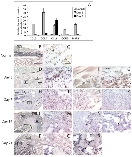
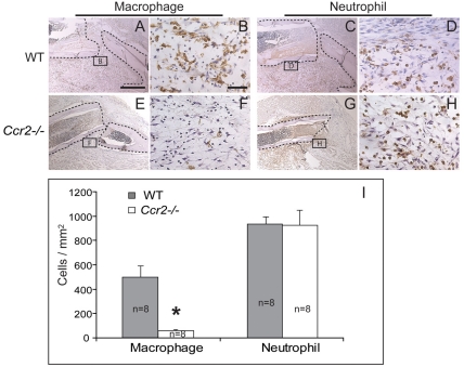
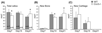
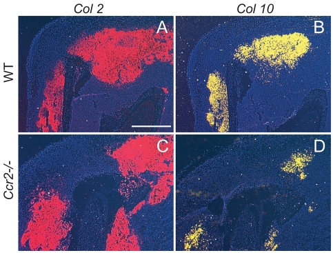
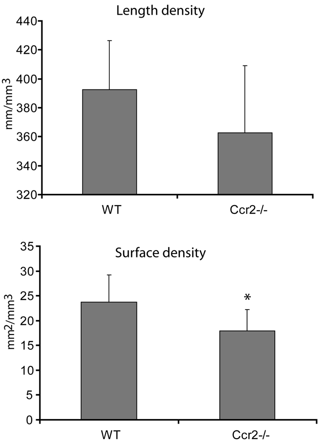
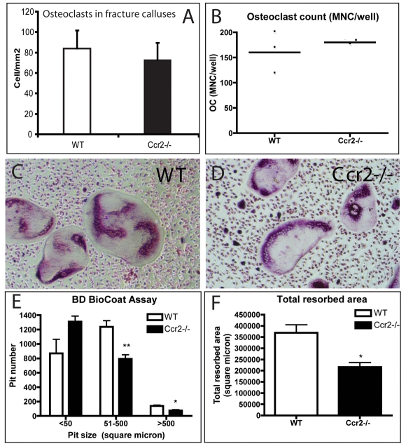
Bone injury induces an inflammatory response that involves neutrophils, macrophages and other inflammatory cells. The recruitment of inflammatory cells to sites of injury occurs in response to specific signaling pathways. The CC chemokine receptor type 2 (CCR2) is crucial for recruiting macrophages, as well as regulating osteoclast function. In this study, we examined fracture healing in Ccr2-/- mice. We first demonstrated that the expression of Ccr2 transcripts and the filtration of macrophages into fracture calluses were most robust during the early phases of fracture healing. We then determined that the number of macrophages at the fracture site was significantly lower in Ccr2-/- mice compared with wild-type controls at 3 days after injury. As a result, impaired vascularization, decreased formation of callus, and delayed maturation of cartilage were observed at 7 days after injury in mutant mice. At day 14, Ccr2-/- mice had less bone in their calluses. At day 21, Ccr2-/- mice had larger calluses and more bone compared with wild-type mice, suggesting a delayed remodeling. In addition, we examined the effect of Ccr2 mutation on osteoclasts. We found that a lack of Ccr2 did not affect the number of osteoclasts within fracture calluses at 21 days after injury. However, Ccr2-/- osteoclasts exhibited a decreased ability to resorb bone compared with wild-type cells, which could contribute to the delayed remodeling of fracture calluses observed in Ccr2-/- mice. Collectively, these results indicate that a deficiency of Ccr2 reduces the infiltration of macrophages and impairs the function of osteoclasts, leading to delayed fracture healing.


Figures

Fig. 1.
Fig. 1.
Mobilization of macrophages during normal fracture healing. (A) RT-PCR assay of the expression levels of molecules related to macrophage recruitment or endocytosis. CCL2, CCL7 and CCL8 are chemokine (CC motif) ligand 2, 7 and 8, respectively. CCR2 is CC chemokine receptor type 2. MSR1 is macrophage scavenger receptor 1. (B,C) In normal unfractured tibiae, macrophages are detected in subcutaneous tissues. Macrophages are stained brown after immunohistochemistry using anti-F4/80 antibody. (D) At 3 days after fracture, abundant macrophages are present in granulation tissues (E), between injured muscles (F), and in the region of periosteal reaction (G). (H) At 7 days after fracture, macrophages are observed in fibrous callus (I) and between newly formed trabecular bone (J), but not in cartilage (K). (L) At day 14, macrophages are present at the periphery of fracture callus (M). A small number of macrophages were also detected at the front of endochondral ossification (N, arrow) and in the new bone (O, arrow). (P) At day 21, macrophages are observed at the periphery of callus (Q). (R)Macrophages were also detected lining trabecular bone in the callus (arrows). The dashed lines in B, D, H, L and P show fracture ends. The dashed line in G shows cortical bone. Bars, 1 mm (B,D,H,L,P); 50 μm (C,E–G,I–K,M–O,Q,R).
Fig. 2.
Fig. 2.
Quantification of macrophages and neutrophils at the fracture sites at 3 days after injury. Macrophages were detected by using the anti-F4/80 antibody. Neutrophils were detected with an MCA771G antibody. Macrophages (A,B) and neutrophils (C,D) are abundant at the fracture site in wild-type mice (WT). (E,F) In Ccr2−/− mice, very few macrophages are present at the fracture site. (G,H) Abundant neutrophils are detected in mutant mice. (I) Further quantitative analysis confirms that the number of macrophages, but not neutrophils, is significantly lower in mutant mice than wild-type mice. Dashed lines in A, C, E and G show fracture ends. *P<0.01. Bars, 1 mm (A,C,E,G); 50 μm (B,D,F,H).
Fig. 3.
Fig. 3.
Histomorphometric analysis of fracture callus in Ccr2−/− mice. (A) The callus size is smaller at day 7, but larger at day 21, in Ccr2−/− mice compared with wild-type mice. (B) The volume of new bone is smaller at day 14, but larger at day 21, in Ccr2−/− mice compared with wild-type mice. (C) There is no difference in the volume of new cartilage between these two groups at all time points. Data in graphs are mean ± standard deviation (S.D.). *P<0.05.
Fig. 4.
Fig. 4.
Delayed chondrocyte maturation in Ccr2−/− mice. (A,B) In situ hybridization shows that Col2 (A) and Col10 (B) transcripts are present in the fracture callus of wild-type mice at day 7. (C,D) In Ccr2−/− mice, abundant Col2 expression is observed at this time (C), but Col10 expression is greatly reduced compared with controls (D). Bar, 1 mm.
Fig. 5.
Fig. 5.
Quantification of blood vessels in the callus at day 7 after fracture. There is no difference in the length density of blood vessels between Ccr2−/− mice and wild-type mice. The surface density of blood vessels in the fracture callus is significantly lower in Ccr2−/− mice than that in the wild-type mice. Data in graphs are mean ± S.D. *P<0.05.
Fig. 6.
Fig. 6.
Osteoclast number and function. (A) There is no significant difference in the number of osteoclasts in fracture calluses of Ccr2−/− mice and wild-type mice at day 21, and (B) the differentiation of osteoclasts in vitro is not altered in the mutants. (C,D)Micrographs of the pits created in discs by wild-type (C) and mutant (D) osteoclasts. (E,F) Ccr2−/− osteoclasts create smaller numbers of larger pits (E) and resorb less surface area (F) in vitro than wild-type osteoclasts. MNC refers to multinucleated cells. *P<0.05, **P<0.01.

References

    1. Altman RD, Latta LL, Keer R, Renfree K, Hornicek FJ, Banovac K. (1995). Effect of nonsteroidal antiinflammatory drugs on fracture healing: a laboratory study in rats. J Orthop Trauma 9, 392–400 - PubMed
    1. Binder NB, Niederreiter B, Hoffmann O, Stange R, Pap T, Stulnig TM, Mack M, Erben RG, Smolen JS, Redlich K. (2009). Estrogen-dependent and C-C chemokine receptor-2-dependent pathways determine osteoclast behavior in osteoporosis. Nat Med. 15, 417–424 - PubMed
    1. Bonnarens F, Einhorn TA. (1984). Production of a standard closed fracture in laboratory animal bone. J Orthop Res. 2, 97–101 - PubMed
    1. Boring L, Gosling J, Chensue SW, Kunkel SL, Farese RV, Jr, Broxmeyer HE, Charo IF. (1997). Impaired monocyte migration and reduced type 1 (Th1) cytokine responses in C-C chemokine receptor 2 knockout mice. J Clin Invest. 100, 2552–2261 - PMC - PubMed
    1. Capoccia BJ, Gregory AD, Link DC. (2008). Recruitment of the inflammatory subset of monocytes to sites of ischemia induces angiogenesis in a monocyte chemoattractant protein-1-dependent fashion. J Leukoc Biol. 84, 760–768 - PMC - PubMed

Publication types

Substances