

Bone marrow-derived endothelial progenitor cells and endothelial cells may contribute to endothelial repair in the kidney immediately after ischemia-reperfusion
- PMID: 20354148
- PMCID: PMC2907274
- DOI: 10.1369/jhc.2010.956011
Bone marrow-derived endothelial progenitor cells and endothelial cells may contribute to endothelial repair in the kidney immediately after ischemia-reperfusion
Abstract
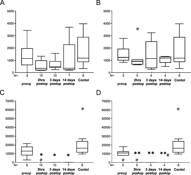
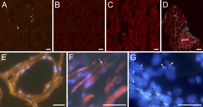
In ischemic acute kidney injury, renal blood flow is decreased. We have previously shown that reperfused, transplanted kidneys exhibited ischemic injury to vascular endothelium and that preservation of peritubular capillary endothelial integrity may be critical to recovery from ischemic injury. We hypothesized that bone marrow-derived (BMD) endothelial progenitor cells (EPCs) might play an important role in renal functional recovery after ischemia. We tested this hypothesis in recipients of cadaveric renal allografts before and for 2 weeks after transplantation. We found that the numbers of circulating CD34-positive EPCs and CD146-positive endothelial cells (ECs) decreased immediately after ischemia-reperfusion. In renal allograft tissues obtained 1 hr after reperfusion, CD34-positive cells were more frequently observed along the endothelial lining of peritubular capillaries compared with non-ischemic controls. Moreover, 0-17.5% of peritubular capillary ECs were of recipient origin. In contrast, only 0.1-0.7% of tubule cells were of recipient origin. Repeat graft biopsy samples obtained 35 and 73 days after transplant did not contain capillary ECs of recipient origin, whereas 1.4% and 12.1% of tubule cells, respectively, were of recipient origin. These findings suggest that BMD EPCs and ECs may contribute to endothelial repair immediately after ischemia-reperfusion.
Figures
Similar articles
-
Preservation of peritubular capillary endothelial integrity and increasing pericytes may be critical to recovery from postischemic acute kidney injury.Am J Physiol Renal Physiol. 2008 Aug;295(2):F351-9. doi: 10.1152/ajprenal.90276.2008. Epub 2008 Jun 18. Am J Physiol Renal Physiol. 2008. PMID: 18562634 Free PMC article.
-
Effects of ischemic preconditioning in the late phase on homing of endothelial progenitor cells in renal ischemia/reperfusion injury.Transplant Proc. 2013 Mar;45(2):511-6. doi: 10.1016/j.transproceed.2012.05.095. Transplant Proc. 2013. PMID: 23498786
-
Presence of endothelial progenitor cells, distinct from mature endothelial cells, within human CD146+ blood cells.Thromb Haemost. 2005 Dec;94(6):1270-9. doi: 10.1160/TH05-07-0499. Thromb Haemost. 2005. PMID: 16411405
-
Microvasculopathy in ischemic acute kidney injury: consequences and therapeutic implications.Panminerva Med. 2012 Mar;54(1):45-52. Panminerva Med. 2012. PMID: 22278116 Review.
-
[The role of endothelial progenitor cells in renal disease].G Ital Nefrol. 2008 Sep-Oct;25(5):537-46. G Ital Nefrol. 2008. PMID: 18828116 Review. Italian.
Cited by
-
Stem/progenitor cell in kidney: characteristics, homing, coordination, and maintenance.Stem Cell Res Ther. 2021 Mar 20;12(1):197. doi: 10.1186/s13287-021-02266-0. Stem Cell Res Ther. 2021. PMID: 33743826 Free PMC article. Review.
-
Bone marrow-derived cells homing for self-repair of periodontal tissues: a histological characterization and expression analysis.Int J Clin Exp Pathol. 2015 Oct 1;8(10):12379-89. eCollection 2015. Int J Clin Exp Pathol. 2015. PMID: 26722424 Free PMC article.
-
Extrarenal Progenitor Cells Do Not Contribute to Renal Endothelial Repair.J Am Soc Nephrol. 2016 Jun;27(6):1714-26. doi: 10.1681/ASN.2015030321. Epub 2015 Oct 9. J Am Soc Nephrol. 2016. PMID: 26453608 Free PMC article.
-
Detection of intrathrombotic endothelial progenitor cells and its application to thrombus age estimation in a murine deep vein thrombosis model.Int J Legal Med. 2017 Nov;131(6):1633-1638. doi: 10.1007/s00414-017-1668-5. Epub 2017 Aug 21. Int J Legal Med. 2017. PMID: 28828642
-
Endothelial colony-forming cells and pro-angiogenic cells: clarifying definitions and their potential role in mitigating acute kidney injury.Acta Physiol (Oxf). 2018 Feb;222(2):10.1111/apha.12914. doi: 10.1111/apha.12914. Epub 2017 Jul 25. Acta Physiol (Oxf). 2018. PMID: 28656611 Free PMC article. Review.
References
-
- Ali T, Khan I, Simpson W, Prescott G, Townend J, Smith W, MacLeod A (2007) Incidence and outcomes in acute kidney injury: a comprehensive population-based study. J Am Soc Nephrol 18:1292–1298 - PubMed
-
- Arendshorst WJ, Finn WF, Gottschalk CW (1976) Micropuncture study of acute renal failure following temporary renal ischemia in the rat. Kidney Int Suppl 6(suppl):100–105 - PubMed
-
- Asahara T, Murohara T, Sullivan A, Silver M, van der Zee R, Li T, Witzenbichler B, et al. (1997) Isolation of putative progenitor endothelial cells for angiogenesis. Science 275:964–967 - PubMed
-
- Becherucci F, Mazzinghi B, Ronconi E, Peired A, Lazzeri E, Sagrinati C, Romagnani P, et al. (2009) The role of endothelial progenitor cells in acute kidney injury. Blood Purif 27:261–270 - PubMed
Publication types
MeSH terms
Substances
LinkOut - more resources
Full Text Sources
Medical
Molecular Biology Databases
