Review
. 2010 May;119(5):523-41.
doi: 10.1007/s00401-010-0679-9. Epub 2010 Mar 31.

Intraneuronal beta-amyloid accumulation and synapse pathology in Alzheimer's disease

Affiliations
Review

Intraneuronal beta-amyloid accumulation and synapse pathology in Alzheimer's disease

Gunnar K Gouras et al. Acta Neuropathol. 2010 May.

Abstract

The aberrant accumulation of aggregated beta-amyloid peptides (Abeta) as plaques is a hallmark of Alzheimer's disease (AD) neuropathology and reduction of Abeta has become a leading direction of emerging experimental therapies for the disease. The mechanism(s) whereby Abeta is involved in the pathophysiology of the disease remain(s) poorly understood. Initially fibrils, and subsequently oligomers of extracellular Abeta have been viewed as the most important pathogenic form of Abeta in AD. More recently, the intraneuronal accumulation of Abeta has been described in the brain, although technical considerations and its relevance in AD have made this a controversial topic. Here, we review the emerging evidence linking intraneuronal Abeta accumulation to the development of synaptic pathology and plaques in AD, and discuss the implications of intraneuronal beta-amyloid for AD pathology, biology, diagnosis and therapy.


Conflict of interest statement

Conflict of interest statement: The authors declare that they have no commercial or financial relationship that could be construed as a potential conflict of interest.

Figures

Fig. 1
Fig. 1
Schematic diagram of APP and APP metabolites, including APP βCTF and Aβ, cleavage sites in APP of α, β and γ-secretases, and domains recognized by representative antibodies. Aβ/APP antibodies, such as 6E10 and 4G8, recognize full-length APP, CTFs and Aβ. Antibodies P2-1 and 369 are directed against the N- and C-terminal regions of full-length APP, respectively. Antibody 3D6 is specific to the free N-terminus of Aβ1-x and βCTF; this antibody will not recognize full-length APP or other APP CTFs. Antibodies MBC40 and MBC42 specifically recognize the free C-terminus of Aβ40 and Aβ42, respectively, and do not react against full-length APP or APP CTFs.
Fig. 2
Fig. 2
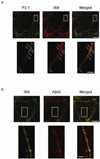
Confocal microscopy of endogenous APP, APP CTFs and Aβ in human primary neurons in culture (ScienCell; 12 days in vitro). a. Dual-immunofluorescence with N-terminal APP antibody P2-1 (green, Affinity BioReagents) and C-terminal APP antibody 369 (red). Lower panels: high power images show full-length APP (arrows) co-labeled by both N- and C-terminal APP antibodies in distal neurites. Next to full-length APP, antibody 369 also recognizes various APP CTFs and the APP intracellular domain (AICD), which is a reason for why antibody 369 gives a more diffuse pattern of staining (central panel) compared to the more punctate labeling with the N-terminal APP antibody P2-1 alone (left panel). Scale bars: 50 µm (upper panels); 10 µm (lower panels). b. Dual-immunofluorescence with antibody 369 (green) and Aβ42 antibody 12F4 (red, Covance). Lower panels: high power images show distinct patterns of staining for antibodies 369 (left panel) and Aβ42 (central panel), with no substantial co-localization (right panel). Antibody 369 labels full-length APP, various APP CTFs and AICD, while the Aβ42-specific antibody labels only Aβx-42 peptides. Scale bars: 25 µm (upper panels); 5 µm (lower panels).
Fig. 3
Fig. 3
Schematic representation of Aβ42 accumulation within neurites leading to Aβ plaque formation in AD. Image at left is a low power representation of neurons with dendritic arborizations in the brain with early Aβ42 accumulation; the red arrow indicates the area of a higher power view shown in the image at right. In the higher power view, accumulation of intraneuronal Aβ42 (red) is associated with altered morphology of these distal neurites and synapses. Immuno-histochemical and –EM studies in AD transgenic mouse and human brains support that aberrant Aβ42 accumulation in dendritic arborizations can be important determinants for the morphology of plaques.
Fig. 4
Fig. 4
Intraneuronal Aβ42 accumulation with head injury. a. Stereotactic insertion of a needle into the brain of a young wild-type rat as a model of head injury. Increased levels of Aβ42 can be seen in CA1 pyramidal neurons and their apical dendrites located near (red line) the needle track (arrow) compared to neurons located further away (blue line) 48 to 72 hours following brain injury. b. Immuno-gold staining demonstrated Aβ42 accumulation particularly within endosomes (arrows) of CA1 pyramidal neurons located near to the needle track. c. Dual-immuno-staining for Aβ42 and AT8 (hyperphosphorylated tau) by light microscopy revealed Aβ42 positive neurons (pink, black arrow), AT8 positive neurons (blue, white arrow) and also co-localization of Aβ42 and AT8 within neurons (purple, arrow head) in postmortem brain from a case with dementia pugilistica. CA1: CA1 pyramidal cell body layer, SR: stratum radiatum. Scale bars: 100 µm (a, c); 2µm (b).

Similar articles

Cited by

References

    1. Alafuzoff I, Pikkarainen M, Arzberger T, et al. Inter-laboratory comparison of neuropathological assessments of beta-amyloid protein: a study of the BrainNet Europe consortium. Acta Neuropathol. 2008;115:533–546. - PubMed
    1. Almeida CG, Takahashi RH, Gouras GK. Beta-amyloid accumulation impairs multivesicular body sorting by inhibiting the ubiquitin-proteasome system. J Neurosci. 2006;26:4277–4288. - PMC - PubMed
    1. Almeida CG, Tampellini D, Takahashi RH, et al. Beta-amyloid accumulation in APP mutant neurons reduces PSD-95 and GluR1 in synapses. Neurobiol Dis. 2005;20:187–198. - PubMed
    1. Allsop D, Haga S, Bruton C, Ishii T, Roberts GW. Neurofibrillary tangles in some cases of dementia pugilistica share antigens with amyloid beta-protein of Alzheimer's disease. Am J Pathol. 1990;136:255–260. - PMC - PubMed
    1. Aoki M, Volkmann I, Tjernberg LO, Winblad B, Bogdanovic N. Amyloid betapeptide levels in laser capture microdissected cornu ammonis 1 pyramidal neurons of Alzheimer's brain. Neuroreport. 2008;19:1085–1089. - PubMed

Publication types