

An affinity matured minibody for PET imaging of prostate stem cell antigen (PSCA)-expressing tumors
- PMID: 20354850
- PMCID: PMC2903645
- DOI: 10.1007/s00259-010-1433-1
An affinity matured minibody for PET imaging of prostate stem cell antigen (PSCA)-expressing tumors
Abstract
Purpose: Prostate stem cell antigen (PSCA), a cell surface glycoprotein expressed in normal human prostate and bladder, is over-expressed in the majority of localized prostate cancer and most bone metastases. We have previously shown that the hu1G8 minibody, a humanized anti-PSCA antibody fragment (single-chain Fv-C(H)3 dimer, 80 kDa), can localize specifically and image PSCA-expressing xenografts at 21 h post-injection. However, the humanization and antibody fragment reformatting decreased its apparent affinity. Here, we sought to evaluate PET imaging contrast with affinity matured minibodies.
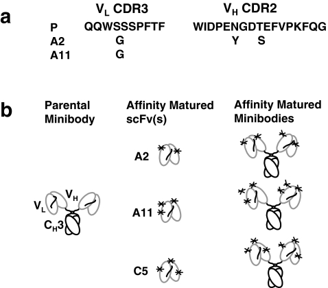
Methods: Yeast scFv display, involving four rounds of selection, was used to generate the three affinity matured antibody fragments (A2, A11, and C5) that were reformatted into minibodies. These three affinity matured anti-PSCA minibodies were characterized in vitro, and following radiolabeling with (124)I were evaluated in vivo for microPET imaging of PSCA-expressing tumors.
Results: The A2, A11, and C5 minibody variants all demonstrated improved affinity compared to the parental (P) minibody and were ranked as follows: A2 > A11 > C5 > P. The (124)I-labeled A11 minibody demonstrated higher immunoreactivity than the parental minibody and also achieved the best microPET imaging contrast in two xenograft models, LAPC-9 (prostate cancer) and Capan-1 (pancreatic cancer), when evaluated in vivo.
Conclusion: Of the affinity variant minibodies tested, the A11 minibody that ranked second in affinity was selected as the best immunoPET tracer to image PSCA-expressing xenografts. This candidate is currently under development for evaluation in a pilot clinical imaging study.
Figures


Similar articles
-
Applications of immunoPET: using 124I-anti-PSCA A11 minibody for imaging disease progression and response to therapy in mouse xenograft models of prostate cancer.Clin Cancer Res. 2014 Dec 15;20(24):6367-78. doi: 10.1158/1078-0432.CCR-14-1452. Epub 2014 Oct 17. Clin Cancer Res. 2014. PMID: 25326233 Free PMC article.
-
124I-Labeled anti-prostate stem cell antigen affinity-matured A11 minibody.2010 Jun 23 [updated 2010 Jul 19]. In: Molecular Imaging and Contrast Agent Database (MICAD) [Internet]. Bethesda (MD): National Center for Biotechnology Information (US); 2004–2013. 2010 Jun 23 [updated 2010 Jul 19]. In: Molecular Imaging and Contrast Agent Database (MICAD) [Internet]. Bethesda (MD): National Center for Biotechnology Information (US); 2004–2013. PMID: 20662135 Free Books & Documents. Review.
-
Quantitative immunoPET of prostate cancer xenografts with 89Zr- and 124I-labeled anti-PSCA A11 minibody.J Nucl Med. 2014 Mar;55(3):452-9. doi: 10.2967/jnumed.113.120873. Epub 2014 Feb 6. J Nucl Med. 2014. PMID: 24504052 Free PMC article.
-
Humanized radioiodinated minibody for imaging of prostate stem cell antigen-expressing tumors.Clin Cancer Res. 2008 Nov 15;14(22):7488-96. doi: 10.1158/1078-0432.CCR-07-5093. Clin Cancer Res. 2008. PMID: 19010866 Free PMC article.
-
124I-Anti-PSCA 2B3 minibody.2009 Mar 25 [updated 2009 May 13]. In: Molecular Imaging and Contrast Agent Database (MICAD) [Internet]. Bethesda (MD): National Center for Biotechnology Information (US); 2004–2013. 2009 Mar 25 [updated 2009 May 13]. In: Molecular Imaging and Contrast Agent Database (MICAD) [Internet]. Bethesda (MD): National Center for Biotechnology Information (US); 2004–2013. PMID: 20641706 Free Books & Documents. Review.
Cited by
-
Molecular imaging of prostate cancer: translating molecular biology approaches into the clinical realm.Eur Radiol. 2015 May;25(5):1294-302. doi: 10.1007/s00330-014-3539-5. Epub 2015 Feb 20. Eur Radiol. 2015. PMID: 25693661 Free PMC article. Review.
-
ImmunoPET of Malignant and Normal B Cells with 89Zr- and 124I-Labeled Obinutuzumab Antibody Fragments Reveals Differential CD20 Internalization In Vivo.Clin Cancer Res. 2017 Dec 1;23(23):7242-7252. doi: 10.1158/1078-0432.CCR-17-0855. Epub 2017 Sep 19. Clin Cancer Res. 2017. PMID: 28928164 Free PMC article.
-
On-demand radiosynthesis of N-succinimidyl-4-[18F]fluorobenzoate ([18F]SFB) on an electrowetting-on-dielectric microfluidic chip for 18F-labeling of protein.RSC Adv. 2019 Oct 9;9(55):32175-32183. doi: 10.1039/c9ra06158d. eCollection 2019 Oct 7. RSC Adv. 2019. PMID: 35530758 Free PMC article.
-
Targeted Radionuclide Therapy of Prostate Cancer-From Basic Research to Clinical Perspectives.Molecules. 2020 Apr 10;25(7):1743. doi: 10.3390/molecules25071743. Molecules. 2020. PMID: 32290196 Free PMC article. Review.
-
Engineered antibodies for molecular imaging of cancer.Methods. 2014 Jan 1;65(1):139-47. doi: 10.1016/j.ymeth.2013.09.015. Epub 2013 Oct 1. Methods. 2014. PMID: 24091005 Free PMC article. Review.
References
-
- Seltzer MA, Barbaric Z, Belldegrun A, Naitoh J, Dorey F, Phelps ME, et al. Comparison of helical computerized tomography, positron emission tomography and monoclonal antibody scans for evaluation of lymph node metastases in patients with prostate specific antigen relapse after treatment for localized prostate cancer. J Urol. 1999;162:1322–1328. doi: 10.1016/S0022-5347(05)68277-8. - DOI - PubMed
Publication types
MeSH terms
Substances
Grants and funding
LinkOut - more resources
Full Text Sources
Other Literature Sources
Medical
Miscellaneous
