Review
. 2010 Apr 7;16(13):1567-76.
doi: 10.3748/wjg.v16.i13.1567.

Diffusion weighted imaging in the liver

Affiliations
Review

Diffusion weighted imaging in the liver

Petra G Kele et al. World J Gastroenterol. .

Abstract

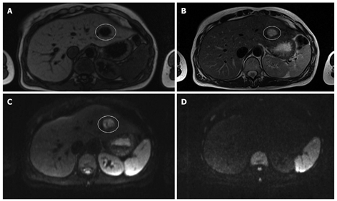
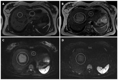
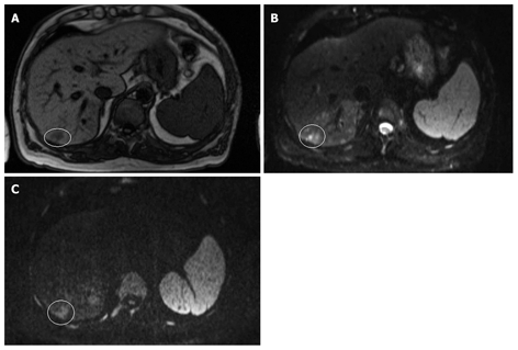
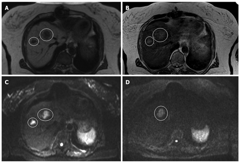
Diffusion weighted magnetic resonance imaging (DWI) is an imaging technique which provides tissue contrast by the measurement of diffusion properties of water molecules within tissues. Diffusion is expressed in an apparent diffusion coefficient (ADC), which reflects the diffusion properties unique to each type of tissue. DWI has been originally used in neuroradiology. More recently, DWI has increasingly been used in addition to conventional unenhanced and enhanced magnetic resonance imaging (MRI) in other parts of the body. The reason for this delay was a number of technical problems inherent to the technique, making DWI very sensitive to artifacts, which had to be overcome. With assessment of ADC values, DWI proved to be helpful in characterization of focal liver lesions. However, DWI should always be used in conjunction to conventional MRI since there is considerable overlap between ADC values of benign and malignant lesions. DWI is useful in the detection of hepatocellular carcinoma in the cirrhotic liver and detection of liver metastases in oncological patients. In addition, DWI is a promising tool in the prediction of tumor responsiveness to chemotherapy and the follow-up of oncological patients after treatment, as DWI may be capable of detecting recurrent disease earlier than conventional imaging. This review focuses on the most common applications of DWI in the liver.


Figures

Figure 1
Figure 1
Brownian movements in hypocellular (A) and hypercellular (B) environment. A: Tissue with low cellularity permits movement of the water molecules; B: Tissue with high cellularity restricts the movement of water molecules.
Figure 2
Figure 2
Magnetic resonance imaging (MRI) and diffusion weighted imaging (DWI) of a cyst. A: T1-weighted MRI; B: T2-weighted MRI; C: Diffusion weighted image (b-value 50 s/mm2); D: Diffusion weighted image (b-value 1000 s/mm2) in a 33-year old woman with multiple liver lesions. The cyst is hypo-intense on the T1-weighted image, hyper-intense on the T2-weighted image and the diffusion weighted image at a b-value 50 s/mm2. Note that the cyst totally disappears on the diffusion weighted image at a b-value 1000 s/mm2.
Figure 3
Figure 3
MRI and DWI of an adenoma and a hemangioma. A: T1-weighted MRI; B: T2-weighted MRI; C: Diffusion weighted image (b-value 50 s/mm2); D: Diffusion weighted image (b-value 1000 s/mm2) in a 41-year-old women with multiple liver lesions. The large lesion in segment 8 is an adenoma and the small one in segment 2-3 is a hemangioma. On the T1-weighted image, the adenoma is slightly hypo-intense to the normal liver parenchyma, and hyper-intense on the T2-weighted image. On the diffusion weighted images, it remains hyper-intense at both b-values. The hemangioma is also hypo-intense on the T1-weighted image and hyper-intense on the T2-weighted image. However, in contrast to the adenoma, it totally disappears at a b-value of 1000 mm/s2.
Figure 4
Figure 4
MRI and DWI of hepatocellular carcinoma. A: T1-weighted; B: DWI (b-value 50 s/mm2); C: Diffusion weighted image (b-value 1000 s/mm2) in a 67-year-old male with hemophilia, hepatitis C-based liver cirrhosis and HCC in segment 7. The HCC is hypo-intense on the T1-weighted image and hyper-intense on the diffusion weighted images at both b-values. Note that the lesion remains hyper-intense on the image with a b-value 1000 s/mm2.
Figure 5
Figure 5
MRI and DWI of hepatic metastases. A: T1-weighted; B: T2-weighted; C: Diffusion weighted image (b-value 50 s/mm2); D: Diffusion weighted image (b-value 1000 s/mm2) in a 74-year-old woman with a history of rectal cancer, recently diagnosed with lung and liver metastases in segment 4 and 8, respectively. The lesions are hypo-intense to the liver parenchyma on the T1-weighted image and hyper-intense on the T2-weighted image. On the diffusion weighted image at a b-value 50 s/mm2, both lesions appear hyper-intense, but at a b-value 1000 s/mm2, only the lesion in segment 4 remains hyper-intense. The lesion in segment 8 has completely disappeared.

Similar articles

Cited by

References

    1. Bammer R. Basic principles of diffusion-weighted imaging. Eur J Radiol. 2003;45:169–184. - PubMed
    1. Naganawa S, Kawai H, Fukatsu H, Sakurai Y, Aoki I, Miura S, Mimura T, Kanazawa H, Ishigaki T. Diffusion-weighted imaging of the liver: technical challenges and prospects for the future. Magn Reson Med Sci. 2005;4:175–186. - PubMed
    1. Charles-Edwards EM, deSouza NM. Diffusion-weighted magnetic resonance imaging and its application to cancer. Cancer Imaging. 2006;6:135–143. - PMC - PubMed
    1. Thoeny HC, De Keyzer F. Extracranial applications of diffusion-weighted magnetic resonance imaging. Eur Radiol. 2007;17:1385–1393. - PubMed
    1. Kwee TC, Takahara T, Ochiai R, Nievelstein RA, Luijten PR. Diffusion-weighted whole-body imaging with background body signal suppression (DWIBS): features and potential applications in oncology. Eur Radiol. 2008;18:1937–1952. - PMC - PubMed