Case Reports
. 2010 Jan;16(1):20-8.
doi: 10.1111/j.1601-0825.2009.01593.x.

Utility of PDL progenitors for in vivo tissue regeneration: a report of 3 cases

Affiliations
Case Reports

Utility of PDL progenitors for in vivo tissue regeneration: a report of 3 cases

F Feng et al. Oral Dis. 2010 Jan.

Abstract

Objective: Periodontal disease is an inflammatory disorder with widespread morbidities involving both oral and systemic health. The primary goal of periodontal treatment is the regeneration of the lost or diseased periodontium. In this study, we retrospectively examined feasibility and safety of reconstructing the periodontal intrabony defects with autologous periodontal ligament progenitor (PDLP) implantation in three patients.

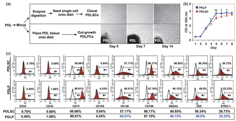
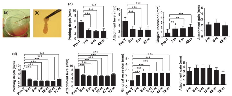
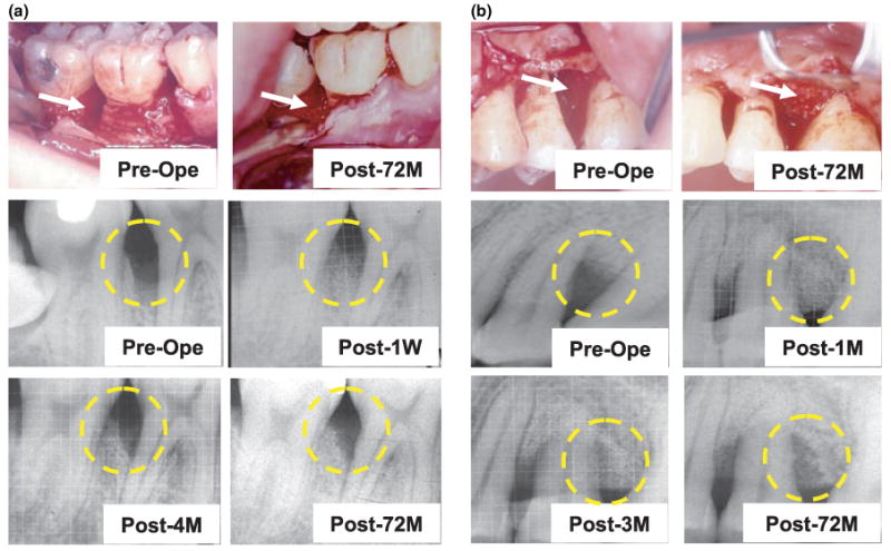
Materials and methods: In this retrospective pilot study, we treated 16 teeth with at least one deep intrabony defect of probing depth (PD) > OR = 6 mm with PDLP transplantation and evaluated clinical outcome measures in terms of probing depth, gingival recession and attachment gain for a duration of 32-72 months. Furthermore, we compare PDLPs with standard PDL stem cells (PDLSCs) and confirmed that PDLPs possessed progenitor characters.

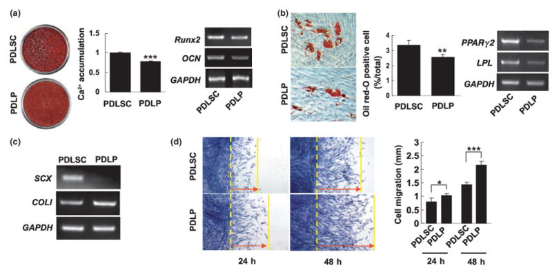
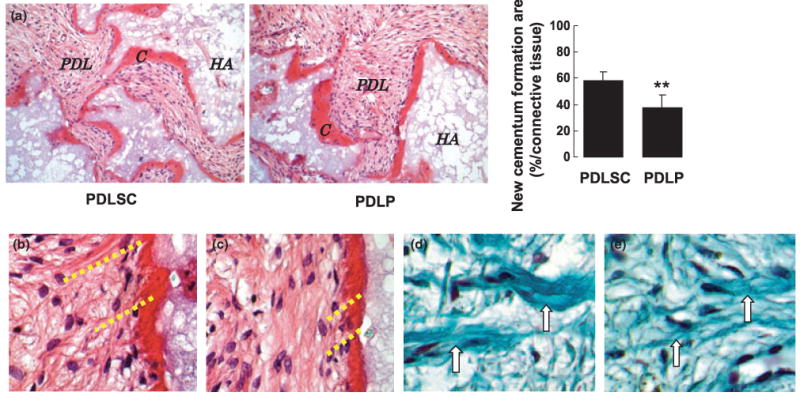
Results: Clinical examination indicated that transplantationof PDLPs may provide therapeutic benefit for the periodontal defects. All treated patients showed no adverse effects during the entire course of follow up. We also found that PDLPs were analogous to PDLSCs in terms of high proliferation, expression of mesenchymal surface molecules, multipotent differentiation, and in vivo tissue regain. However, PDLPs failed to express scleraxis, a marker of tendon, as seen in PDLSCs.

Conclusions: This study demonstrated clinical and experimental evidences supporting a potential efficacy and safety of utilizing autologous PDL cells in the treatment of human periodontitis.


Figures

Figure 1
Figure 1
Isolation of human PDLPs. (a) Comparison procedure of isolating PDLPs and PDLSCs from periodontal tissues and morphology of culture expanded cells at 5, 7, and 14 days (magnification 100×). (b) At days 1–3, PDLPs showed similar proliferation rate as PDLSCs, but showed an elevated proliferation from day 4 to 7. After day 8, PDLPs have similar proliferation rate as PDLSCs in the culture. (c) Cytometric flow analysis indicated that PDLPs have similar surface molecule phenotype as PDLSCs derived from same donor including negative for CD34 and CD45, and positive for CD73, CD105, CD146, CD166, SSEA4, 3G5, and STRO-1 (*P < 0.05; **P < 0.01). Experiments were repeated three times
Figure 2
Figure 2
Comparison differentiation of PDLPs with PDLSCs. (a) PDLPs were cultured with L-ascorbate-2-phosphate, dexamethasone, and inorganic phosphate for 4 weeks. Alizarin red staining showed that PDLPs form mineralized nodules. In comparison with induced PDLSCs, PDLP cultures accumulated significantly less amounts of calcium (P < 0.005) than that of PDLSCs. PDLPs expressed Runx2 and OCN by RT-PCR analysis, but expression levels were lower than PDLSCs. (b) Under the adipogenic induction, PDLPs were capable of forming oil red-O positive cells, but PDLPs form significantly less oil red-O cells than PDLSCs (P < 0.01) (magnification 100×). RT-PCR analysis showed that PDLPs express lower levels of PPARγ2 and LPL when compared with PDLSCs. (c) RT-PCR analysis showed that cultured PDLPs failed to express SCX, a transcription factor specifically expressed in tendon cells and PDLSCs. But analogous to PDLSCs, PDLPs express COLI. (d) Cell Scratch assay showed that PDLPs have significantly higher cell motility than PDLSCs at 24 and 48 h (*P < 0.05; ***P < 0.005) (magnification 40×). Experiments were repeated three times
Figure 3
Figure 3
PDLPs generated cementum-like and PDL-like structures in vivo. (a) After 8 weeks of transplantation, PDLPs were able to form cementum-like structure (C) on the surfaces of (HA) carrier, PDL-like tissue (PDL) was also generated. But PDLPs generated significantly less amount of cementum than PDLSCs (**P < 0.01) (magnification 200×). (b, c) H&E staining showed that PDLPs were capable of regenerating Sharpey's fibers (b) as seen in PDLSC transplant (c) (magnification 400×). (d, e) Trichrome staining showed that PDLP transplants contain newly regenerated collagen fibers (d, arrows) same as observed in PDLSC transplants (e, arrows) (magnification 400×)
Figure 4
Figure 4
Autologous PDLP-mediated treatment to periodontitis. (a, b) Procedures of generating PDLPs/bone grafting material CALCITITE 4060-2 complex. Autologous PDLPs were cultured with bone grafting material CALCITITE 4060-2 and reach confluence. After washing with PBS, PDLP/bone grafting material CALCITITE 4060-2 complex was removed (a) and delivered to periodontal defect area in which inflammatory tissues were surgically removed (b). (c) In patient no. 1, 12 teeth were treated and followed up for 3, 6, and 42 months. From 3 to 42 month post-treatment, there was significantly decreased probing depths and attachment losses compared with the pretreatment level (Pre-T) (***P < 0.001). Although there was significantly increased gingival recession compared with the pretreatment (Pre-T) (***P < 0.001, **P < 0.005), attachment gain also marked increased after treatment. Moreover, there was no significant change of attachment gain on 42 months after treatment compared with that of on 3 months. (d) In patient no. 2, three teeth were treated and followed up for 3, 6, 9, 12, 42 and 72 months. From 3 to 72 month post-treatment, there was significantly decreased probing depths and attachment losses compared with the pretreatment level (Pre-T) (***P < 0.001). There was significantly increased gingival recession compared with the pretreatment (Pre-T) (***P < 0.001), however, attachment gain also marked increased after treatment. There was no significant change of attachment gain among the different time points after treatment
Figure 5
Figure 5
Clinical evaluation of autologous PDLP-mediated treatment to periodontitis. (a) Patient no. 1 showed periodontal tissue destruction at preoperation caused by periodontitis in tooth no. 30 (arrow in Pre-Ope). After 72 months autologous PDLP implantation, surgical reentry to the implanted area showed periodontal tissue regeneration (arrows in Post-72M). X-ray analysis showed bone resorption at preoperation (yellow circle in Pre-Ope) and PDLP/carrier material implantation at 1 week postoperation (yellow circle in Post-1W), 4 months postoperation (yellow circle in Post-4M), and 72 months postoperation (yellow circle in Post-72M). (b) Patient no. 2 showed periodontal tissue destruction at preoperation caused by periodontitis in tooth no. 13 (arrow in Pre-Ope). After 72 months autologous PDLP implantation, surgical reentry to the implanted area showed periodontal tissue regeneration (arrows in Post-72M). X-ray analysis showed bone resorption at preoperation (yellow circle in Pre-Ope) and PDLP/carrier material implantation at 1 month postoperation (yellow circle in Post-1M), 3 months postoperation (yellow circle in Post-3M), and 72 months postoperation (yellow circle in Post-72M)

References

    1. Andriankaja OM, Genco RJ, Dorn J, et al. The use of different measurements and definitions of periodontal disease in the study of the association between periodontal disease and risk of myocardial infarction. J Periodontol. 2006;77:1067–1073. - PubMed
    1. Bi Y, Ehirchiou D, Kilts TM, et al. Identification of tendon stem/progenitor cells and their extracellular matrix-rich niche. Nat Med. 2007;13:1219–1227. - PubMed
    1. D'Errico JA, Ouyang H, Berry JE, et al. Immortalized cementoblasts and periodontal ligament cells in culture. Bone. 1999;25:39–47. - PubMed
    1. Geismar K, Stoltze K, Sigurd B, et al. Periodontal disease and coronary heart disease. J Periodontol. 2006;77:1547–1554. - PubMed
    1. Goncalves PF, Gurgel BC, Pimentel SP, et al. Effect of two different approaches for root decontamination on new cementum formation following guided tissue regeneration: a histomorphometric study in dogs. J Periodontal Res. 2006;41:535–540. - PubMed

Publication types