

Near-infrared phosphorescent polymeric nanomicelles: efficient optical probes for tumor imaging and detection
- PMID: 20355951
- PMCID: PMC3681954
- DOI: 10.1021/am9001293
Near-infrared phosphorescent polymeric nanomicelles: efficient optical probes for tumor imaging and detection
Abstract
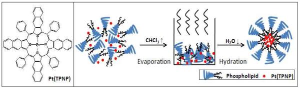
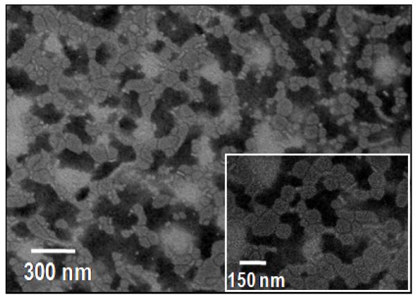
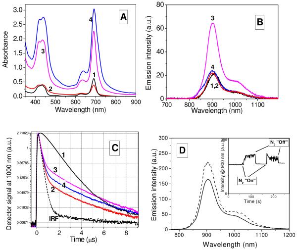
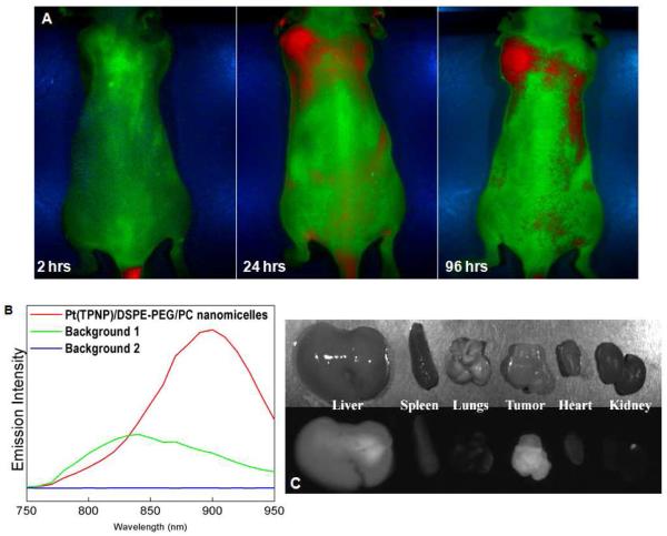
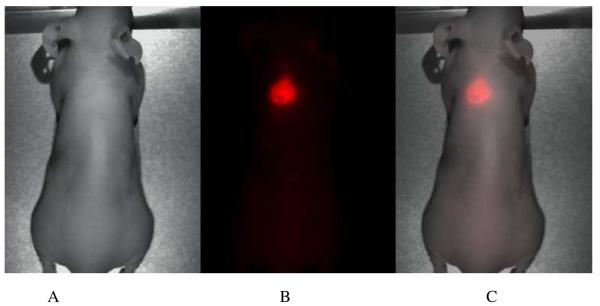
We report a formulation of near-infrared (near-IR) phosphorescent polymeric nanomicelles and their use for in vivo high-contrast optical imaging, targeting, and detection of tumors in small animals. Near-IR phosphorescent molecules of Pt(II)-tetraphenyltetranaphthoporphyrin (Pt(TPNP)) were found to maintain their near-IR phosphorescence properties when encapsulated into phospholipid nanomicelles. The prepared phosphorescent micelles are of approximately 100 nm size and are highly stable in aqueous suspensions. A large spectral separation between the Pt(TPNP) absorption, with a peak at approximately 700 nm, and its phosphorescence emission, with a peak at approximately 900 nm, allows a dramatic decrease in the level of background autofluorescence and scattered excitation light in the near-IR spectral range, where the signal from the phosphorescent probe is observed. In vivo animal imaging with subcutaneously xenografted tumor-bearing mice has resulted in high contrast optical images, indicating highly specific accumulation of the phosphorescent micelles into tumors. Using optical imaging with near-IR phosphorescent nanomicelles, detection of smaller, visually undetectable tumors has also been demonstrated.
Figures
Similar articles
-
Combined magnetic resonance and optical imaging of head and neck tumor xenografts using Gadolinium-labelled phosphorescent polymeric nanomicelles.Head Neck Oncol. 2010 Nov 26;2:35. doi: 10.1186/1758-3284-2-35. Head Neck Oncol. 2010. PMID: 21110873 Free PMC article.
-
Turning double hydrophilic into amphiphilic: IR825-conjugated polymeric nanomicelles for near-infrared fluorescence imaging-guided photothermal cancer therapy.Nanoscale. 2018 Jan 25;10(4):2115-2127. doi: 10.1039/c7nr07495f. Nanoscale. 2018. PMID: 29326993
-
Extending Hypochlorite Sensing from Cells to Elesclomol-Treated Tumors in Vivo by Using a Near-Infrared Dual-Phosphorescent Nanoprobe.ACS Appl Mater Interfaces. 2018 Oct 24;10(42):35838-35846. doi: 10.1021/acsami.8b14717. Epub 2018 Oct 15. ACS Appl Mater Interfaces. 2018. PMID: 30260621
-
Aggregation-enhanced fluorescence in PEGylated phospholipid nanomicelles for in vivo imaging.Biomaterials. 2011 Sep;32(25):5880-8. doi: 10.1016/j.biomaterials.2011.04.080. Epub 2011 May 20. Biomaterials. 2011. PMID: 21601279
-
Emerging applications of phosphorescent metalloporphyrins.J Fluoresc. 2005 Jul;15(4):569-84. doi: 10.1007/s10895-005-2830-x. J Fluoresc. 2005. PMID: 16167215 Review.
Cited by
-
Multifunctional Phosphorescent Conjugated Polymer Dots for Hypoxia Imaging and Photodynamic Therapy of Cancer Cells.Adv Sci (Weinh). 2015 Sep 10;3(2):1500155. doi: 10.1002/advs.201500155. eCollection 2016 Feb. Adv Sci (Weinh). 2015. PMID: 27722081 Free PMC article.
-
Crystallization induced room-temperature phosphorescence and chiral photoluminescence properties of phosphoramides.Chem Sci. 2022 Apr 20;13(20):5893-5901. doi: 10.1039/d2sc00990k. eCollection 2022 May 25. Chem Sci. 2022. PMID: 35685799 Free PMC article.
-
Amphiphilic BODIPY-Hydroporphyrin Energy Transfer Arrays with Broadly Tunable Absorption and Deep Red/Near-Infrared Emission in Aqueous Micelles.J Org Chem. 2017 Jun 16;82(12):6054-6070. doi: 10.1021/acs.joc.7b00357. Epub 2017 Jun 5. J Org Chem. 2017. PMID: 28516773 Free PMC article.
-
BODIPY-Bacteriochlorin Energy Transfer Arrays: Toward Near-IR Emitters with Broadly Tunable, Multiple Absorption Bands.J Org Chem. 2017 Dec 15;82(24):13068-13075. doi: 10.1021/acs.joc.7b02031. Epub 2017 Nov 22. J Org Chem. 2017. PMID: 29119786 Free PMC article.
-
In vivo cancer imaging by poly(ethylene glycol)-b-poly(ɛ-caprolactone) micelles containing a near-infrared probe.Nanomedicine. 2012 Feb;8(2):228-36. doi: 10.1016/j.nano.2011.06.009. Epub 2011 Jun 24. Nanomedicine. 2012. PMID: 21704593 Free PMC article.
References
Publication types
MeSH terms
Substances
Grants and funding
LinkOut - more resources
Full Text Sources
Other Literature Sources
