

Phylogeny and expression of carbonic anhydrase-related proteins
- PMID: 20356370
- PMCID: PMC2873310
- DOI: 10.1186/1471-2199-11-25
Phylogeny and expression of carbonic anhydrase-related proteins
Abstract
Background: Carbonic anhydrases (CAs) are found in many organisms, in which they contribute to several important biological processes. The vertebrate alpha-CA family consists of 16 subfamilies, three of which (VIII, X and XI) consist of acatalytic proteins. These are named carbonic anhydrase related proteins (CARPs), and their inactivity is due to absence of one or more Zn-binding histidine residues. In this study, we analyzed and evaluated the distribution of genes encoding CARPs in different organisms using bioinformatic methods, and studied their expression in mouse tissues using immunohistochemistry and real-time quantitative PCR.
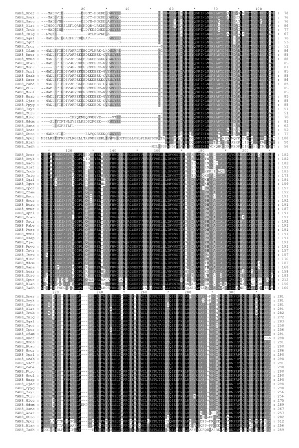
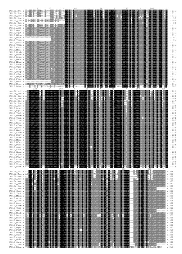
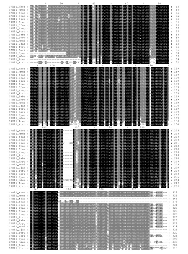
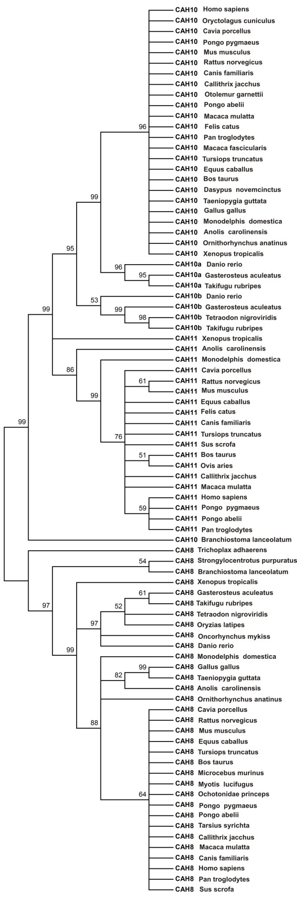
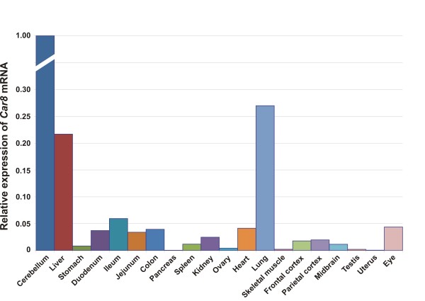
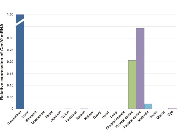
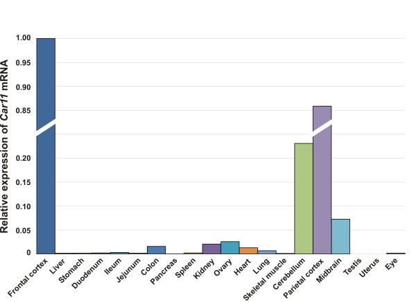
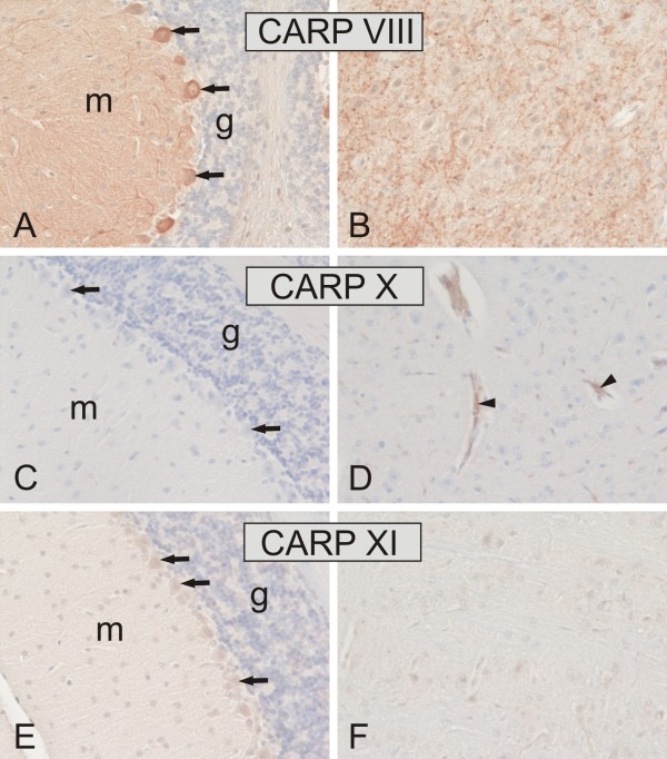
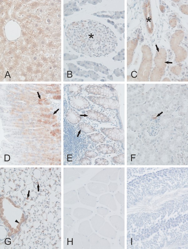
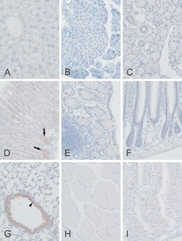
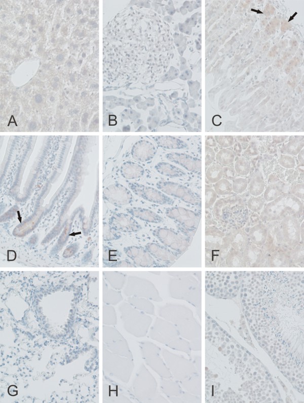
Results: We collected 84 sequences, of which 22 came from novel or improved gene models which we created from genome data. The distribution of CARP VIII covers vertebrates and deuterostomes, and CARP X appears to be universal in the animal kingdom. CA10-like genes have had a separate history of duplications in the tetrapod and fish lineages. Our phylogenetic analysis showed that duplication of CA10 into CA11 has occurred only in tetrapods (found in mammals, frogs, and lizards), whereas an independent duplication of CA10 was found in fishes. We suggest the name CA10b for the second fish isoform. Immunohistochemical analysis showed a high expression level of CARP VIII in the mouse cerebellum, cerebrum, and also moderate expression in the lung, liver, salivary gland, and stomach. These results also demonstrated low expression in the colon, kidney, and Langerhans islets. CARP X was moderately expressed in the cerebral capillaries and the lung and very weakly in the stomach and heart. Positive signals for CARP XI were observed in the cerebellum, cerebrum, liver, stomach, small intestine, colon, kidney, and testis. In addition, the results of real-time quantitative PCR confirmed a wide distribution for the Car8 and Car11 mRNAs, whereas the expression of the Car10 mRNA was restricted to the frontal cortex, parietal cortex, cerebellum, midbrain, and eye.
Conclusions: CARP sequences have been strongly conserved between different species, and all three CARPs show high expression in the mouse brain and CARP VIII is also expressed in several other tissues. These findings suggest an important functional role for these proteins in mammals.
Figures
Similar articles
-
Carbonic anhydrase related proteins: molecular biology and evolution.Subcell Biochem. 2014;75:135-56. doi: 10.1007/978-94-007-7359-2_8. Subcell Biochem. 2014. PMID: 24146378 Review.
-
Evolutionarily conserved, "acatalytic" carbonic anhydrase-related protein XI contains a sequence motif present in the neuropeptide sauvagine: the human CA-RP XI gene (CA11) is embedded between the secretor gene cluster and the DBP gene at 19q13.3.Genomics. 1998 Dec 15;54(3):484-93. doi: 10.1006/geno.1998.5585. Genomics. 1998. PMID: 9878252
-
Sequence of the pearl oyster carbonic anhydrase-related protein and its evolutionary implications.Biochem Genet. 2012 Apr;50(3-4):269-76. doi: 10.1007/s10528-011-9469-x. Epub 2011 Oct 1. Biochem Genet. 2012. PMID: 21964518
-
An update on carbonic anhydrase-related proteins VIII, X and XI.J Enzyme Inhib Med Chem. 2013 Dec;28(6):1129-42. doi: 10.3109/14756366.2012.727813. Epub 2013 Jan 7. J Enzyme Inhib Med Chem. 2013. PMID: 23294106 Review.
-
Carbonic anhydrase related protein VIII and its role in neurodegeneration and cancer.Curr Pharm Des. 2010;16(29):3264-76. doi: 10.2174/138161210793429823. Curr Pharm Des. 2010. PMID: 20819067 Review.
Cited by
-
Expression of the CHOP-inducible carbonic anhydrase CAVI-b is required for BDNF-mediated protection from hypoxia.Brain Res. 2014 Jan 16;1543:28-37. doi: 10.1016/j.brainres.2013.11.018. Epub 2013 Nov 23. Brain Res. 2014. PMID: 24275196 Free PMC article.
-
Calcareous sponge genomes reveal complex evolution of α-carbonic anhydrases and two key biomineralization enzymes.BMC Evol Biol. 2014 Nov 25;14:230. doi: 10.1186/s12862-014-0230-z. BMC Evol Biol. 2014. PMID: 25421146 Free PMC article.
-
Structure based exploration of mitochondrial alpha carbonic anhydrase inhibitors as potential leads for anti-obesity drug development.Daru. 2024 Dec;32(2):907-924. doi: 10.1007/s40199-024-00535-w. Epub 2024 Sep 14. Daru. 2024. PMID: 39276204 Review.
-
Cross study transcriptomic investigation of Alzheimer's brain tissue discoveries and limitations.Sci Rep. 2025 May 8;15(1):16041. doi: 10.1038/s41598-025-01017-y. Sci Rep. 2025. PMID: 40341634 Free PMC article.
-
Exploration of Aromatic Hydrazides as Inhibitors of Human Carbonic Anhydrases.Arch Pharm (Weinheim). 2025 Apr;358(4):e202400963. doi: 10.1002/ardp.202400963. Arch Pharm (Weinheim). 2025. PMID: 40165649 Free PMC article.
References
-
- Tashian RE. Genetics of the mammalian carbonic anhydrases. Adv Genet. 1992;30:321–356. full_text. - PubMed
Publication types
MeSH terms
Substances
LinkOut - more resources
Full Text Sources
Research Materials
