. 2010 Mar 31;12(1):20.
doi: 10.1186/1532-429X-12-20.

Oxygenation-sensitive CMR for assessing vasodilator-induced changes of myocardial oxygenation

Affiliations

Oxygenation-sensitive CMR for assessing vasodilator-induced changes of myocardial oxygenation

Matthias Vöhringer et al. J Cardiovasc Magn Reson. .

Abstract

Background: As myocardial oxygenation may serve as a marker for ischemia and microvascular dysfunction, it could be clinically useful to have a non-invasive measure of changes in myocardial oxygenation. However, the impact of induced blood flow changes on oxygenation is not well understood. We used oxygenation-sensitive CMR to assess the relations between myocardial oxygenation and coronary sinus blood oxygen saturation (SvO2) and coronary blood flow in a dog model in which hyperemia was induced by intracoronary administration of vasodilators.

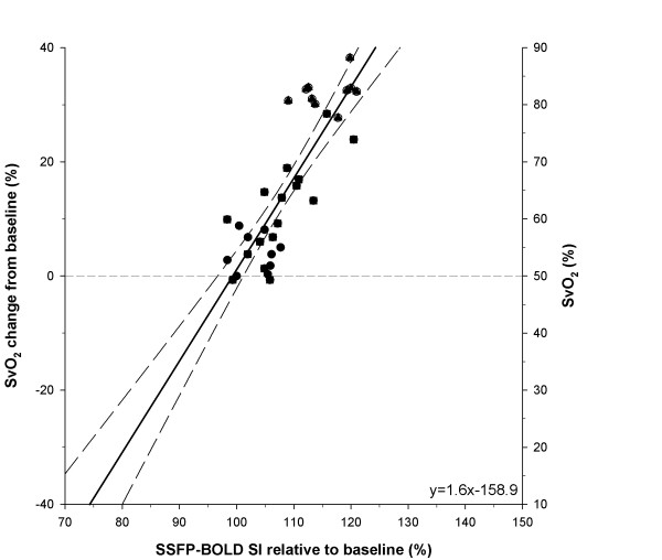
Results: During administration of acetylcholine and adenosine, CMR signal intensity correlated linearly with simultaneously measured SvO2 (r2 = 0.74, P < 0.001). Both SvO2 and CMR signal intensity were exponentially related to coronary blood flow, with SvO2 approaching 87%.

Conclusions: Myocardial oxygenation as assessed with oxygenation-sensitive CMR imaging is linearly related to SvO2 and is exponentially related to vasodilator-induced increases of blood flow. Oxygenation-sensitive CMR may be useful to assess ischemia and microvascular function in patients. Its clinical utility should be evaluated.


Figures

Figure 1
Figure 1
Examples of BOLD-CMR images a) at baseline; c) during infusion of 0.3 mg/min adenosine (Ade 3). The perfusion territory of the LCX after intracoronary injection of gadopentetate dimeglumine is shown in b) (arrows). Panel d) shows the myocardial signal intensity of baseline subtracted from that during adenosine. The higher values in the LCX territory reflect the signal intensity increase during in the LCX perfusion territory as induced by adenosine injection.
Figure 2
Figure 2
Examples of T2* maps at a) baseline, b) during LCX infusion of 10 μg/min acetylcholine (ACh 3) and c) during infusion of 0.3 mg/min adenosine (Ade 3). The perfusion territory of the LCX after intracoronary injection of gadolinium is shown in d) and indicated with white arrows.
Figure 3
Figure 3
Correlation of absolute oxygen saturation (SvO2) in the coronary sinus (CS) and blood flow in the LCX (n = 7). Regression lines (dashed lines) are shown. Baseline, Adenosine and acetylcholine induced changes in flow represented as circles, squares and triangles, respectively. LCX: Left circumflex coronary artery.
Figure 4
Figure 4
Linear correlation of SvO2 changes from baseline values versus myocardial BOLD-CMR signal intensity relative to baseline (n = 6). Regression lines (dashed lines) are shown. Baseline, adenosine and acetylcholine induced changes in flow represented as circles, squares and triangles, respectively. Figure 4 includes the results of the analyses of extrapolated lines. See text. LCX = left circumflex coronary artery.

References

    1. Feigl EO. Coronary physiology. Physiol Rev. 1983;63:1–205. - PubMed
    1. Wilke NM, Jerosch-Herold M, Zenovich A, Stillman AE. Magnetic resonance first-pass myocardial perfusion imaging: clinical validation and future applications. J Magn Reson Imaging. 1999;10:676–685. doi: 10.1002/(SICI)1522-2586(199911)10:5<676::AID-JMRI10>3.0.CO;2-L. - DOI - PubMed
    1. McCommis KS, Goldstein TA, Abendschein DR, Herrero P, Misselwitz B, Gropler RJ, Zheng J. Quantification of regional myocardial oxygenation by magnetic resonance imaging: validation with positron emission tomography. Circ Cardiovasc Imaging. 2010;3:41–46. doi: 10.1161/CIRCIMAGING.109.897546. - DOI - PMC - PubMed
    1. Kennan RP, Zhong J, Gore JC. Intravascular susceptibility contrast mechanisms in tissues. Magn Reson Med. 1994;31:9–21. doi: 10.1002/mrm.1910310103. - DOI - PubMed
    1. Wendland MF, Saeed M, Lauerma K, de Crespigny A, Moseley ME, Higgins CB. Endogenous susceptibility contrast in myocardium during apnea measured using gradient recalled echo planar imaging. Magn Reson Med. 1993;29:273–276. doi: 10.1002/mrm.1910290220. - DOI - PubMed