. 2010 Apr 1:10:86.
doi: 10.1186/1471-2334-10-86.

Changes of tau profiles in brains of the hamsters infected with scrapie strains 263 K or 139 A possibly associated with the alteration of phosphate kinases

Affiliations

Changes of tau profiles in brains of the hamsters infected with scrapie strains 263 K or 139 A possibly associated with the alteration of phosphate kinases

Gui-Rong Wang et al. BMC Infect Dis. .

Abstract

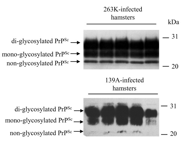
Background: Phospho-tau deposition has been described in a rare genetic human prion disease, Gerstmann-Sträussler-Scheinker syndrome, but is not common neuropathological picture for other human and animal transmissible spongiform encephalopathies (TSEs). This study investigated the possible changes of tau and phosphorylated tau (p-tau, at Ser396, Ser404, and Ser202/Thr205) in scrapie experimental animals.

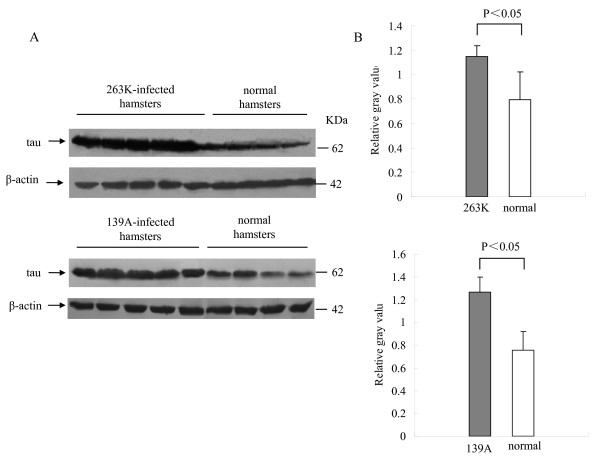
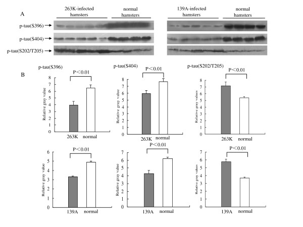
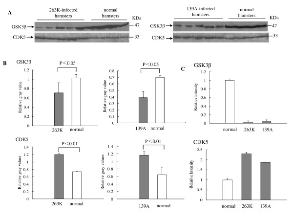
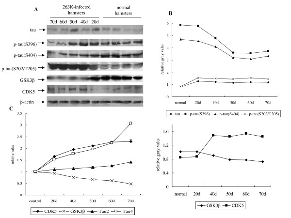
Methods: The profiles of tau and p-tau (p-tau, at Ser396, Ser404, and Ser202/Thr205) in the brain tissues of agents 263K- or 139A-infected hamsters were evaluated by Western blots and real-time PCR. Meanwhile, the transcriptional and expressive levels of GSK3beta and CDK5 in the brains were tested.

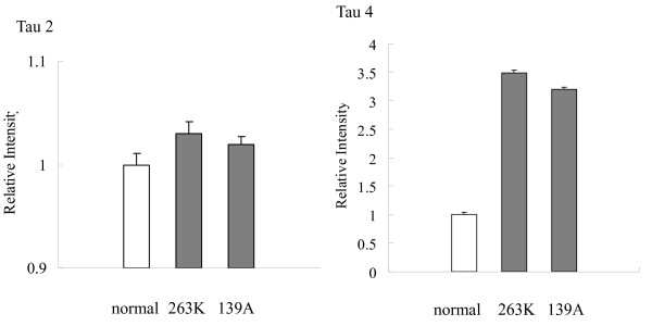
Results: The contents of total tau and p-tau at Ser202/Thr205 increased, but p-tau at Ser396 and Ser404 decreased at the terminal stages, regardless of scrapie strains. Transcriptional levels of two tau isoforms were also increased. Additionally, it showed higher CDK5, but lower GSK3beta transcriptional and expressive levels in the brains of scrapie-infected animals. Analysis of brain samples collected from different times after inoculated with agent 263 K revealed that the changes of tau profiles and phosphate kinases were time-relative events.

Conclusion: These data suggest that changes of profiles of p-tau at Ser396, Ser404 and Ser202/Thr205 are illness-correlative phenomena in TSEs, which may arise of the alteration of phosphate kinases. Alteration of tau, p-tau (Ser396, Ser404, and Ser202/Thr205), GSK3beta and CDK5 were either intermediate or consequent events in TSE pathogenesis and proposed the potential linkage of these bioactive proteins with the pathogenesis of prion diseases.


Figures

Figure 1
Figure 1
Western blot analysis of PrPSc in the brain tissues of scrapie agents 263 K- and 139A-infected hamsters collected at the moribund stage. For detection of PrPSc, all tested brain homogenates were digested with a final concentration of 50 μg/ml PK at 37°C for 60 min. Same amounts of individual brain homogenate were loaded in 15% SDS-PAGE. The scrapie agents were shown on the top of the graphs. PrPSc specific immunoblots (non-, mono- and di-glycosylated PrP isoforms) were indicated by arrows on the left and molecular mass markers were indicated to the right.
Figure 2
Figure 2
Comparative analysis of the level of tau protein in the brain tissues of normal, scrapie agent 263K- and 139A-infected hamsters collected at the moribund stage. A. Western blots. Same amounts of individual brain homogenate were loaded in 12% SDS-PAGE. The scrapie agents as well as normal control were shown on the top of the graphs. Tau specific immunoblots were indicated by arrows on the left and molecular mass markers were indicated to the right. B. Quantitative analyses of each gray numerical value of tau vs that of β-actin. The average values were calculated from five individual infected hamsters or four individual normal hamsters and presented as mean ± SD. Statistical differences compared with normal controls were illustrated as P < 0.05 and P < 0.01.
Figure 3
Figure 3
Comparative analysis of the mRNA transcription of Tau2 and Tau4 in the brain tissues of normal and scrapie-infected hamsters collected at the moribund stage by real time PCR. The transcription level of each specific mRNA was determined relative to that of the individual β-actin. The relative intensity of each gene from scrapie-infected hamsters was relative to that of respective gene from normal hamsters that was set to 1. Data are representative of three independent experiments.
Figure 4
Figure 4
Analysis of the levels of p-tau (Ser396), p-tau (Ser404) and p-tau (Ser202/Thr205) in the brain tissues of normal and scrapie-infected hamsters collected at the moribund stage. A. Western blots. Same amounts of individual brain homogenate were loaded in 12% SDS-PAGE. The scrapie agents and normal controls were shown on the top of the graphs. Various specific immunoblots were indicated by arrows on the left. B. Quantitative analysis of each gray numerical value of p-tau (Ser396), p-tau (Ser404) and p-tau (Ser202/Thr205) vs that of β-actin. The average values were calculated from five individual infected hamsters or four individual normal hamsters and presented as mean ± SD. Statistical differences compared with controls were illustrated as P < 0.05 and P < 0.01.
Figure 5
Figure 5
Analysis of GSK3β and CDK5 in the brain tissues of normal and scrapie-infected hamsters collected at the moribund stage. A. Western blots. Same amounts of individual brain homogenate were loaded in 12% SDS-PAGE. The scrapie agents and normal controls were shown on the top of the graphs. Various specific immunoblots were indicated by arrows on the left. B. Quantitative analysis of each gray numerical value of GSK3β and CDK5 vs that of β-actin. The average values were calculated from five individual infected hamsters or four individual normal hamsters and presented as mean ± SD. Statistical differences compared with controls were illustrated as P < 0.05 and P < 0.01. C. Real time PCR analysis of the mRNA transcription of GSK3β and CDK5. The transcription level of each specific mRNA was determined relative to that of the individual β-actin. The relative intensity of each gene from scrapie-infected hamsters was relative to that of respective gene from normal hamsters that was set to 1. Data are representative of three independent experiments.
Figure 6
Figure 6
Dynamic analyses of tau, p-tau (Ser396), p-tau (Ser404), p-tau (Ser202/Thr205), GSK3β and CDK5 in the brain tissues of normal and scrapie 263K-infected hamsters during incubation period. A. Western blots. Same amounts of individual brain homogenate were loaded in 12% SDS-PAGE. The scrapie agent 263 K and normal controls were shown on the top of the graphs. Various specific immunoblots were indicated by arrows on the left. B. Quantitative analysis of each gray numerical value of tau, p-tau (Ser396), p-tau (Ser404), p-tau (Ser202/Thr205), GSK3β and CDK5 vs that of β-actin. The average values were calculated from two individual infected hamsters or four individual normal hamsters and presented as mean ± SD. Statistical differences compared with controls were illustrated as P < 0.05 and P < 0.01. C. Real time PCR analysis of the mRNA transcription of Tau2, Tau4, GSK3β and CDK5. The transcription level of each specific mRNA was determined relative to that of the individual β-actin. The relative intensity of each gene from scrapie-infected hamsters was relative to that of respective gene from normal hamsters that was set to 1. Data are representative of three independent experiments.

Similar articles

Cited by

References

    1. Prusiner SB. Prions. Proc Natl Acad Sci. 1998;95:13363–13383. doi: 10.1073/pnas.95.23.13363. - DOI - PMC - PubMed
    1. Collinge J. Prion diseases of humans and animals: their causes and molecular basis. Annu Rev Neurosci. 2001;24:519–550. doi: 10.1146/annurev.neuro.24.1.519. - DOI - PubMed
    1. Goedert M, Spillantini MG, Jakes R, Rutherford D, Crowther RA. Multiple isoforms of human microtubule-associated protein tau: sequences and localization in neurofibrillary tangles of Alzheimer's disease. Neuron. 1989;3:519–526. doi: 10.1016/0896-6273(89)90210-9. - DOI - PubMed
    1. Chun W, Johnson GV. The role of tau phosphorylation and cleavage in neuronal cell death. Front Biosci. 2007;12:733–756. doi: 10.2741/2097. - DOI - PubMed
    1. Goedert M. Tau protein and the neurofibrillary pathology of Alzheimer's disease. Trends Neurosci. 1993;16:460–465. doi: 10.1016/0166-2236(93)90078-Z. - DOI - PubMed

Publication types

LinkOut - more resources