. 2010 Jun;31(18):4889-97.
doi: 10.1016/j.biomaterials.2010.02.062. Epub 2010 Mar 30.

The initiation of embryonic-like collagen fibrillogenesis by adult human tendon fibroblasts when cultured under tension

Affiliations

The initiation of embryonic-like collagen fibrillogenesis by adult human tendon fibroblasts when cultured under tension

Monika L Bayer et al. Biomaterials. 2010 Jun.

Abstract

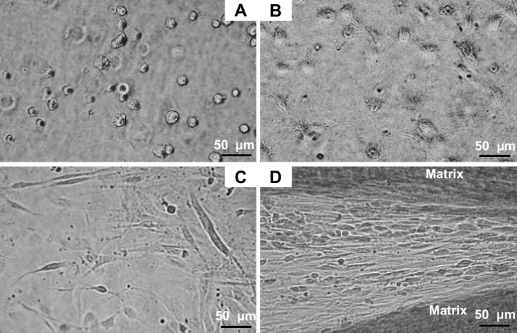
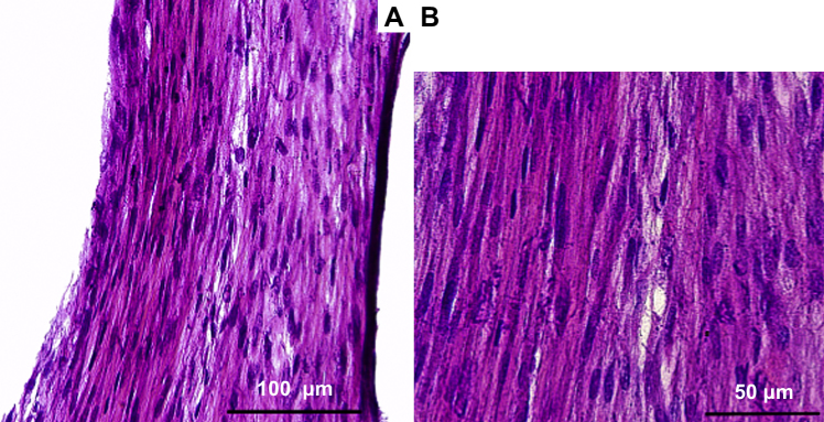
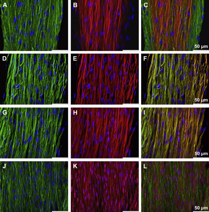
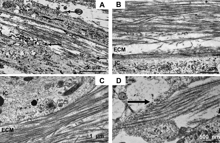
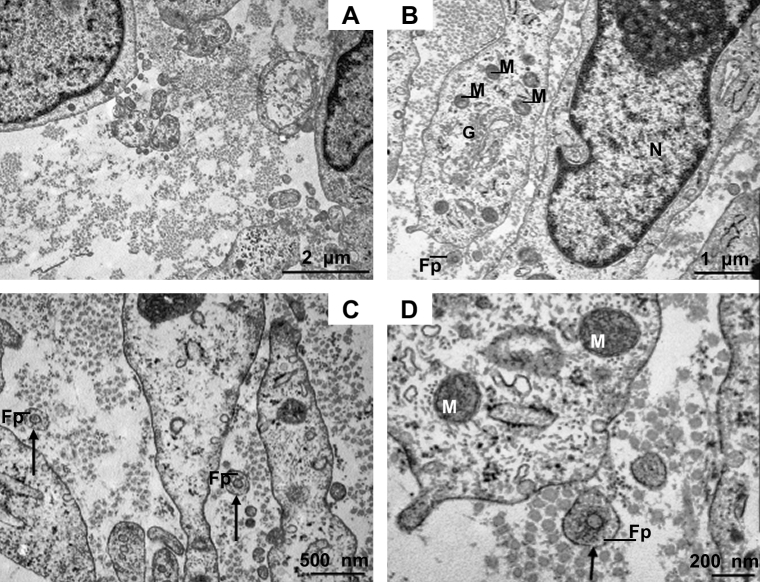
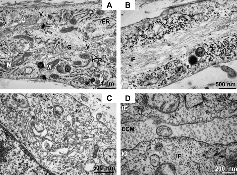
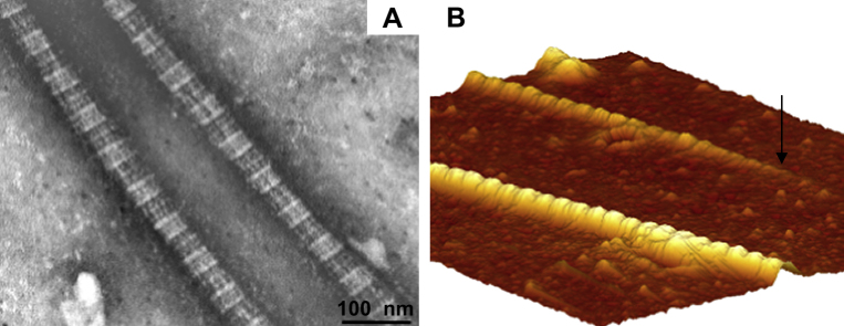
Tendon fibroblasts synthesize collagen and form fibrils during embryonic development, but to what extent mature fibroblasts are able to recapitulate embryonic development and develop normal tendon structure is unknown. The present study examined the capability of mature human tendon fibroblasts to initiate collagen fibrillogenesis when cultured in fixed-length fibrin gels. Fibroblasts were dissected from semitendinosus and gracilis tendons from healthy humans and cultured in 3D linear fibrin gels. The fibroblasts synthesized an extracellular matrix of parallel collagen fibrils that were aligned along the axis of tension. The fibrils had a homogeneous narrow diameter that was similar to collagen fibrils occurring in embryonic tendon. Immunostaining showed colocalization of collagen type I with collagen III, XII and XIV. A fibronectin network was formed in parallel with the collagen, and fibroblasts stained positive for integrin alpha(5). Finally, the presence of cell extensions into the extracellular space with membrane-enclosed fibrils in fibripositors indicated characteristics of embryonic tendon. We conclude that mature human tendon fibroblasts retain an intrinsic capability to perform collagen fibrillogenesis similar to that of developing tendon, which implies that the hormonal/mechanical milieu, rather than intrinsic cellular function, inhibits regenerative potential in mature tendon.


Figures

Fig. 1
Fig. 1
Human tendon fibroblasts embedded in a 3D fibrin gel. (A) Tendon fibroblasts display a rounded cell morphology shortly after embedding in fibrin. The image is taken approximately 15 min following cell seeding. Bar: 50 μm. (B) Fibroblasts are expanding and form cellular extensions 180 min following seeding. Bar: 50 μm. (C) Fibroblasts display a thin, elongated cell body and long extensions, the random alignment is seen in the early phase (first three days) of construct formation. Bar: 50 μm. (D) Parallel arrangement of tendon fibroblasts, which are located in between a synthesized matrix with high cellularity. The ordered alignment is visible during the late stage of construct contraction and tissue formation. Bar: 50 μm.
Fig. 2
Fig. 2
H&E staining of a longitudinal cryosection of a tendon construct. Parallel alignment of fibrils in the extracellular matrix and the dense arrangement of cell nuclei are demonstrated. Bar 2A: 100 μm, Bar 2B: 50 μm.
Fig. 3
Fig. 3
Immunohistological images of tendon constructs. (A) Collagen I (green) and nuclear counterstain DAPI (blue), (B) collagen III (red) and DAPI (blue), (C) computer-generated merged images of the individually captured images. Bar: 50 μm, (D) Collagen I (green) and DAPI (blue), (E) collagen XII (red) and DAPI (blue), (F) computer-generated merged images of the individually captured images. Bar: 50 μm, (G) Collagen I (green) and DAPI (blue), (H) collagen XIV (red) and DAPI (blue), (I) computer-generated merged images of the individually captured images. Bar: 50 μm, (J) Fibronectin (green) and DAPI (blue) (K) integrin α5 (red) and DAPI (blue), (L) computer-generated merged images of the individually captured images. Bar: 50 μm.
Fig. 4
Fig. 4
Transmission electron micrographs of tendon constructs. (A)–(D) longitudinally sectioned micrographs. (A) The image shows the arrangement of collagen fibrils in between two tendon fibroblasts. Arrows point to membrane convolutions. Bar: 5 μm. (B) Parallel alignment of collagen fibrils in the ECM adjacent to a tendon fibroblast. Bar: 1 μm. (C) The electron micrograph displays collagen fibrils in the extracellular space in close proximity to a tendon fibroblast. Bar: 1 μm. (D) Collagen-deposition site on a fibroblast is shown. Bar: 500 nm ECM: extracellular matrix, IC: intracellular space.
Fig. 5
Fig. 5
Transmission electron micrographs of tendon constructs. (A)–(D) transverse sectioned micrographs. (A) The image demonstrates the uniformity of collagen fibrils with a narrow diameter. Bar: 2 μm. (B) Tendon fibroblasts in close proximity and collagen fibrils in the extracellular space. In the intracellular space, mitochondria, the Golgi apparatus and the nucleus can be identified. Bar: 1 μm. (C) Collagen fibrils in the extracellular space are visible and collagen fibrils within fibripositors clearly identifiable. Fibripositors are indicated by arrows. Bar: 500 nm. (D) Collagen fibril in fibripositors at high magnification. Bar: 200 nm. G: Golgi apparatus, Fp: fibripositors, M: mitochondrion, N: nucleus.
Fig. 6
Fig. 6
Electron micrographs of tendon fibroblasts, longitudinally (A, B) and transverse (C, D) sectioned. (A) The electron micrograph displays the intracellular space with cell organelles. The Golgi apparatus is seen in close proximity of multiple vesicles, the cell has formed an extensive network of rough Endoplasmatic Reticulum. Several mitochondria are visible. Bar: 1 μm. (B) Image shows a micrograph with an elaborate regular parallel arrangement of intracellular filaments. Bar: 500 nm. (C) Golgi apparatus with multiple vesicles at high magnification. Bar: 500 nm. (D) The transverse section displays the extensive network of intracellular filaments. Bar: 200 nm. G: Golgi apparatus, IF: intracellular filaments, M: mitochondrion, rER: rough endoplasmatic reticulum, V: vesicle.
Fig. 7
Fig. 7
Electron micrograph and image obtained by Atomic Force Microscopy. (A) The image shows two negatively stained collagen fibrils, demonstrating the regular banding pattern of collagen. Bar: 100 nm (B) 3D reconstruction of an atomic force micrograph, which reveals the characteristic collagen banding pattern and shows a collagen fibril end (indicated by arrow). The original image in 2D has a size of 2 μm × 2 μm.
Fig. 8
Fig. 8
Force-elongation curve of a tendon construct pulled until failure, raw data of three experiments. The curves show the well-described toe and linear region and serves as a test of the tendon constructs’ integrity and mechanical behavior.

Similar articles

Cited by

References

    1. Kastelic J., Galeski A., Baer E. The multicomposite structure of tendon. Connect Tissue Res. 1978;6(1):11–23. - PubMed
    1. Birk D.E., Trelstad R.L. Extracellular compartments in tendon morphogenesis: collagen fibril, bundle, and macroaggregate formation. J Cell Biol. 1986;103(1):231–240. - PMC - PubMed
    1. Canty E.G., Lu Y., Meadows R.S., Shaw M.K., Holmes D.F., Kadler K.E. Coalignment of plasma membrane channels and protrusions (fibripositors) specifies the parallelism of tendon. J Cell Biol. 2004;165(4):553–563. - PMC - PubMed
    1. Humphries S.M., Lu Y., Canty E.G., Kadler K.E. Active negative control of collagen fibrillogenesis in vivo. Intracellular cleavage of the type I procollagen propeptides in tendon fibroblasts without intracellular fibrils. J Biol Chem. 2008;283(18):12129–12135. - PubMed
    1. Richardson S.H., Starborg T., Lu Y., Humphries S.M., Meadows R.S., Kadler K.E. Tendon development requires regulation of cell condensation and cell shape via cadherin-11-mediated cell–cell junctions. Mol Cell Biol. 2007;27(17):6218–6228. - PMC - PubMed

Publication types