. 2010 Jun 11;285(24):18352-63.
doi: 10.1074/jbc.M109.077602. Epub 2010 Mar 31.

Thrombin and collagen induce a feedback inhibitory signaling pathway in platelets involving dissociation of the catalytic subunit of protein kinase A from an NFkappaB-IkappaB complex

Affiliations

Thrombin and collagen induce a feedback inhibitory signaling pathway in platelets involving dissociation of the catalytic subunit of protein kinase A from an NFkappaB-IkappaB complex

Stepan Gambaryan et al. J Biol Chem. .

Abstract

Protein kinase A (PKA) activation by cAMP phosphorylates multiple target proteins in numerous platelet inhibitory pathways that have a very important role in maintaining circulating platelets in a resting state. Here we show that in thrombin- and collagen-stimulated platelets, PKA is activated by cAMP-independent mechanisms involving dissociation of the catalytic subunit of PKA (PKAc) from an NFkappaB-IkappaBalpha-PKAc complex. We demonstrate mRNA and protein expression for most of the NFkappaB family members in platelets. From resting platelets, PKAc was co-immunoprecipitated with IkappaBalpha, and conversely, IkappaBalpha was also co-immunoprecipitated with PKAc. This interaction was significantly reduced in thrombin- and collagen-stimulated platelets. Stimulation of platelets with thrombin- or collagen-activated IKK, at least partly by PI3 kinase-dependent pathways, leading to phosphorylation of IkappaBalpha, disruption of an IkappaBalpha-PKAc complex, and release of free, active PKAc, which phosphorylated VASP and other PKA substrates. IKK inhibitor inhibited thrombin-stimulated IkBalpha phosphorylation, PKA-IkBalpha dissociation, and VASP phosphorylation, and potentiated integrin alphaIIbbeta3 activation and the early phase of platelet aggregation. We conclude that thrombin and collagen not only cause platelet activation but also appear to fine-tune this response by initiating downstream NFkappaB-dependent PKAc activation, as a novel feedback inhibitory signaling mechanism for preventing undesired platelet activation.


Figures
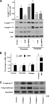
FIGURE 1.
FIGURE 1.
cAMP-independent PKA activation in thrombin- and collagen-stimulated platelets. A–C, washed human platelets (3 × 10⁸/ml) were stimulated for 2 min with thrombin (0.01 unit/ml), collagen (10 μg/ml), or forskolin (0.1 μm), or prior to stimulation preincubated with the indicated inhibitors for 10 min, and then processed for Western blot analysis (A, C) or cAMP/cGMP measurement (B). A, platelets were untreated or preincubated with PKA inhibitors (H-89, 10 μm; Rp-8-pCPT-cAMPS, 200 μm), prior to stimulation with thrombin or forskolin. Platelet activation was monitored by p38 phosphorylation. Immunoblots were scanned and quantified by the Image J program, the intensity of the VASPSer157 signal normalized to the total p38 signal, and then this ratio for each sample was expressed relative to the ratio for thrombin stimulation which was designated as 1. H-89 inhibited both thrombin- and forskolin-stimulated VASPSer157 phosphorylation, whereas Rp-8-pCPT-cAMPS inhibited only forskolin-stimulated VASPSer157 phosphorylation. Results are means ± S.E., n = 4, + p < 0.05 compared with the control; *, p < 0.05 compared with the corresponding stimulus. B, cAMP and cGMP levels in thrombin- and collagen-stimulated platelets. Forskolin- and sodium nitroprusside (SNP) (both 5 μm, 2 min)-stimulated platelets were used as positive controls. cAMP/cGMP concentrations were measured as described under “Experimental Procedures.” cAMP concentration significantly decreased only in thrombin-stimulated platelets. Results are means ± S.E., n = 4, + p < 0.05 compared with control. C, platelets were incubated with thrombin (0.01 unit/ml) or collagen (10 μg/ml), or each of these after preincubation with H89 (10 μm, 10 min) as indicated, and analyzed for VASPSer157 and Rap1GAP2Ser7 phosphorylation, or total Rap1GAP2 expression. Results are representative of three independent experiments.
FIGURE 2.
FIGURE 2.
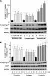
Time and concentration curve of VASPSer157 phosphorylation in thrombin-, collagen-, and forskolin-stimulated platelets. Washed human platelets (3 × 10⁸/ml) were incubated (A) for 1 min with indicated concentrations of thrombin, collagen, or forskolin, or (B) with thrombin (0.01 unit/ml), collagen (1 μg/ml), or forskolin (0.1 μm) for the indicated times, and then processed for Western blot analysis (VASPSer157, total VASP, and GAPDH). Immunoblots were scanned and quantified as described in Fig. 1A. Results are means ± S.E., n = 6, + p < 0.05 compared with no stimulus. Only forskolin-stimulated VASPSer157 phosphorylation was time- and concentration-dependent.
FIGURE 3.
FIGURE 3.
VASPSer157 phosphorylation in thrombin- and convulxin-stimulated platelets is not mediated by PKC, PKB, RhoA kinase, or MEKK/ERK pathways, but may partially involve PI3K. Washed human platelets (3 × 10⁸/ml) were incubated for 1 min with thrombin (0.01 unit/ml), convulxin (5 ng/ml), or forskolin (0.1 μm) as indicated, or each of these after preincubation for 10 min with either (A) PKC inhibitors bisindolylmaleimide I or I× (BisI, BisIX, both 5 μm), (B) wortmannin (0.1–0.5 μm), (C) PKA inhibitor (H-89, 10 μm), or PKB inhibitor (PKI-AKT, 10 μm), (D) PKA inhibitor (H89, 5–20 μm), (E) Rho kinase inhibitor (Y27632, 10 μm), or (F) MEKK1 inhibitor (U0126, 10–50 μm). Results were analyzed by flow cytometry for integrin αIIbβ3 activation (PAC-1 binding) and by Western blotting for VASP, MARCKS, pleckstrin, GSK3, PKB, p38, MLC, and ERK phosphorylation. PAC-1 binding (A, C) represents integrin αIIbβ3 activation and is expressed as % of the thrombin effect, which was designated as 100%. Immunoblots (D) were scanned and the intensity of bands quantified by the Image J program. VASP (□) and PKB (■) phosphorylation were normalized to the total PKB signal. Results are means ± S.E., n = 4. A and C, +significant difference from control; *, significant difference from thrombin- or convulxin- stimulated platelets, respectively. D, +significant difference from thrombin- or forskolin-stimulated platelets, respectively.
FIGURE 4.
FIGURE 4.
Expression of NFκB family proteins and mRNA in platelets. Whole protein lysate and total RNA were prepared from washed platelets and HL60 cells (positive control) and used for Western blotting and RT-PCR analysis as described under “Experimental Procedures.” The same amount of total protein (40 μg/lane) or PCR products (10 μl/lane) from platelet and HL-60 cell lysates was loaded. Presented results are representative of at least three independent experiments.
FIGURE 5.
FIGURE 5.
PKAc is released from an IκB-PKAc complex by thrombin or collagen stimulation of platelets. Washed human platelets (1 × 10⁹/ml) were incubated for 2 min with thrombin (0.01 unit/ml) or collagen (10 μg/ml), pelleted, and resuspended in IP buffer. Specific antibodies were used to precipitate IκBα (A, B, G, H) or PKAc (C, D, G) overnight at 4 °C, followed by protein A or G-Sepharose beads for 1 h (IP). Precipitated proteins were solubilized, separated by SDS-PAGE, and immunoblotted (WB), and blots were treated with IκBα (A, D, G) or PKAc (B, C, G) antibodies. As control, nonspecific antibodies (IgG) were used for IP. In G, platelets were preincubated with or without IKK inhibitor VII (5 μm, 10 min), then stimulated with thrombin (0.01 unit/ml) for 2 min and analyzed by Western blotting for VASP phosphorylation (WB, without prior IP), or analyzed by IP with IκBα or PKAc antibodies and subsequent WB. To estimate the total concentration of PKAc expressed in platelets, in comparison to the amount of PKAc that forms a complex with IκBα, different amount of platelet proteins and purified PKAc were loaded on the same gel as the samples from IP of IκB from platelets (H). Immunoblots were scanned, and the intensity of bands quantified by the Image J program; in E and F, intensities were expressed as fold change with respect to control samples which were designated as 1. Results are means ± S.E., n = 4, + significant difference from respective controls.
FIGURE 6.
FIGURE 6.
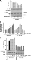
NFκB activation in thrombin-stimulated platelets. A, IκBα phosphorylation and degradation in thrombin-stimulated platelets. Washed human platelets (3 × 10⁸/ml) were incubated with thrombin (0.01 unit/ml, all 6 Western blot panels) for the indicated times, or were first preincubated for 10 min with either 5 μm of IKK inhibitor VII (+IKK inh., third panel from top), or with 10 μm proteasome inhibitor (+MG132, fifth panel from top). Samples were analyzed by Western blotting for VASPSer157 and IκBαSer32/36 phosphorylation, as well as for NFκB and IκBα total protein expression. Immunoblots were scanned, and the intensity of bands was quantified by the Image J program. IκBα was normalized to the NFκB signal, and the ratio was expressed as fold change with respect to the control sample (0 min thrombin), which was designated as 1. B, time-dependent thrombin-induced platelet and HL-60 cell NFκB activation. Washed human platelets (3 × 10⁸/ml) and HL-60 cells (1 × 10⁷ cells/ml) were incubated with thrombin (0.01 unit/ml for platelets, 0.1 unit/ml for HL-60 cells) for the indicated times, then analyzed for NFκB activation using a DNA binding assay (ELISA), as described under “Experimental Procedures.” C, IKK inhibitor inhibits thrombin, but not forskolin-induced VASPSer157 phosphorylation, and potentiates thrombin-stimulated PAC-1 binding. Washed human platelets (3 × 10⁸/ml) were stimulated with thrombin (0.001 unit/ml) or forskolin (1 μm), or preincubated with the indicated concentrations of IKK inhibitor VII for 10 min prior to stimulation, then analyzed by FACS for integrin αIIbβ3 activation (PAC-1 binding), and by Western blotting for VASP and p38 phosphorylation. Results are means ± S.E., n = 3, +, significantly different from control in A and B, and from thrombin in C.
FIGURE 7.
FIGURE 7.
IKK inhibitor potentiates thrombin- and collagen-stimulated platelet aggregation. Washed human platelets (3 × 10⁸/ml) were preincubated with vehicle (0.01% DMSO) or 2 μm IKK inhibitor VII for 5 min. Platelet aggregation was stimulated by addition of 0.01 unit/ml thrombin or 10 μg/ml collagen. A and B, representative aggregation traces showing potentiating effect of IKK inhibitor VII on thrombin (A)- and collagen (B)-induced platelet aggregation. The aggregation in response to thrombin and thrombin + IKK inhibitor VII (C), as well as collagen and collagen + IKK inhibitor VII (D) were calculated as the area under the aggregation curve (AUC, % aggregation × s) and expressed as arbitrary units; aggregation caused by thrombin or collagen alone was designated as 100%. Results are means ± S.E., n = 6, +, significantly different from control in C and D.
FIGURE 8.
FIGURE 8.
Proposed mechanism of cAMP-independent PKAc activation by dissociation of an NFκB-IκB-PKAc complex in thrombin- and collagen- stimulated platelets. In platelets, both collagen and thrombin activate the PI3K pathway that stimulates PKC and PKB which are involved in platelet activation. However, both pathways stimulate IKK and degradation of an NFκB-IκB-PKAc complex, setting free active PKAc that phosphorylates VASP and other substrates involved in platelet inhibitory pathways. Note that in order to simplify the illustration, it was necessary to limit it to the few pathways shown that were the focus of this study. Dotted lines indicate that intermediary agents which transmit thrombin/collagen effects on NFκB-IκB-PKA signaling require further elucidation.

Similar articles

Cited by

References

    1. Italiano J. E., Jr., Patel-Hett S., Hartwig J. H. (2007) J. Thromb. Haemost. 5, Suppl. 1, 18–23 - PubMed
    1. Junt T., Schulze H., Chen Z., Massberg S., Goerge T., Krueger A., Wagner D. D., Graf T., Italiano J. E., Jr., Shivdasani R. A., von Andrian U. H. (2007) Science 317, 1767–1770 - PubMed
    1. Ruggeri Z. M., Mendolicchio G. L. (2007) Circ. Res. 100, 1673–1685 - PubMed
    1. Davì G., Patrono C. (2007) N. Engl. J. Med. 357, 2482–2494 - PubMed
    1. Varga-Szabo D., Pleines I., Nieswandt B. (2008) Arterioscler. Thromb. Vasc. Biol. 28, 403–412 - PubMed

Publication types

LinkOut - more resources