. 2010 Jun 4;285(23):17763-70.
doi: 10.1074/jbc.M109.078980. Epub 2010 Mar 31.

Structural basis for substrate selectivity in human maltase-glucoamylase and sucrase-isomaltase N-terminal domains

Affiliations

Structural basis for substrate selectivity in human maltase-glucoamylase and sucrase-isomaltase N-terminal domains

Lyann Sim et al. J Biol Chem. .

Abstract

Human maltase-glucoamylase (MGAM) and sucrase-isomaltase (SI) are small intestinal enzymes that work concurrently to hydrolyze the mixture of linear alpha-1,4- and branched alpha-1,6-oligosaccharide substrates that typically make up terminal starch digestion products. MGAM and SI are each composed of duplicated catalytic domains, N- and C-terminal, which display overlapping substrate specificities. The N-terminal catalytic domain of human MGAM (ntMGAM) has a preference for short linear alpha-1,4-oligosaccharides, whereas N-terminal SI (ntSI) has a broader specificity for both alpha-1,4- and alpha-1,6-oligosaccharides. Here we present the crystal structure of the human ntSI, in apo form to 3.2 A and in complex with the inhibitor kotalanol to 2.15 A resolution. Structural comparison with the previously solved structure of ntMGAM reveals key active site differences in ntSI, including a narrow hydrophobic +1 subsite, which may account for its additional substrate specificity for alpha-1,6 substrates.


Figures
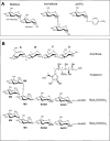
FIGURE 1.
FIGURE 1.
Substrates, inhibitors, and glycan ligands. A, maltose, isomaltose, and pNPG substrates. Non-reducing and reducing ends are indicated for the maltose substrate. B, α-glucosidase inhibitors (acarbose and kotalanol) and Asn822-linked oligosaccharide ligands (Man3GlcNAc2 and Man2GlcNAc2).
FIGURE 2.
FIGURE 2.
Crystal structure and ligands of ntSI. A, arrangement of ntSI monomers A, B, C, and D in the crystal asymmetric unit. B, interaction between monomer B (blue) and monomer C (yellow) observed in crystal-packing lattice and the proposed assembly of the full-length SI protein (composed of O-glycosylated stalk (thick black line) and ntSI and ctSI domains) with respect to the membrane. Approximate locations of the active site pockets are indicated by *. C, representative FoFc electron density maps of ntSI-kotalanol (shown in red mesh) with bound kotalanol (blue) and Man2GlcNAc2 (yellow) and of apo-ntSI (shown as cyan mesh) with bound Man3GlcNAc2 (pink). Maps are contoured at 2.5σ, and Asn822 residues from the ntSI-kotalanol (yellow) and apo-ntSI (pink) structures are shown attached to Man2GlcNAc2 and Man3GlcNAc2, respectively. D, active site superposition of apo-ntSI-monomer C with bound Man3GlcNAc2 glycan (pink) and the ntSI-kotalanol monomer A with bound Man2GlcNAc2 glycan (yellow) and Tris (orange). The Asn822 glycosylation sites attached to the glycan chains are shown as thin pink and yellow sticks, and the catalytic nucleophile Asp472 and acid/base catalyst Asp571 are indicated as thin blue sticks. A double-headed arrow indicates the distance (∼9 Å) between the NAG1 rings of the glycan rings (labeled according to Fig. 1B).
FIGURE 3.
FIGURE 3.
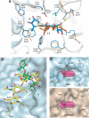
Comparison of ntMGAM and ntSI active sites. For A–C, the approximate locations of the −1 and +1 subsites are labeled. A, superposition of ntMGAM-kotalanol (orange) and ntSI-kotalanol (blue) active sites. Kotalanol and ntMGAM/ntSI residues are represented as thick sticks and thin sticks, respectively. Selected bound waters in the ntMGAM and ntSI structures are shown in yellow and green, respectively. ntMGAM residues are labeled with an asterisk. B, superposition of Man2GlcNAc2 (yellow) and Tris (orange) from ntSI-kotalanol with acarbose (green) from ntMGAM-acarbose in the ntSI active site (blue surface representation). A double-headed arrow indicates the distance (∼7 Å) between the C6-OH of the M4 ring and C1 of acarbose ring A. C, surface representation of the ntSI (top) and ntMGAM (bottom) active sites with non-structurally conserved residues displayed as blue and orange sticks, respectively. One possible conformation of isomaltose (pink, surface-rendered) is modeled into the −1 and +1 subsites.

Similar articles

Cited by

References

    1. Van Beers E. H., Büller H. A., Grand R. J., Einerhand A. W., Dekker J. (1995) Crit. Rev. Biochem. Mol. Biol. 30, 197–262 - PubMed
    1. Semenza G. (1986) Ann. Rev. Cell Biol. 2, 255–313 - PubMed
    1. Nichols B. L., Avery S., Sen P., Swallow D. M., Hahn D., Sterchi E. (2003) Proc. Natl. Acad. Sci. U.S.A. 100, 1432–1437 - PMC - PubMed
    1. Quezada-Calvillo R., Sim L., Ao Z., Hamaker B. R., Quaroni A., Brayer G. D., Sterchi E. E., Robayo-Torres C. C., Rose D. R., Nichols B. L. (2008) J. Nutr. 138, 685–692 - PubMed
    1. Heymann H., Breitmeier D., Günther S. (1995) Biol. Chem. Hoppe Seyler 376, 249–253 - PubMed

Publication types

LinkOut - more resources