. 2010 May 28;285(22):16854-63.
doi: 10.1074/jbc.M110.114546. Epub 2010 Mar 31.

Kruppel-like factor 4 inhibits epithelial-to-mesenchymal transition through regulation of E-cadherin gene expression

Affiliations

Kruppel-like factor 4 inhibits epithelial-to-mesenchymal transition through regulation of E-cadherin gene expression

Jennifer L Yori et al. J Biol Chem. .

Abstract

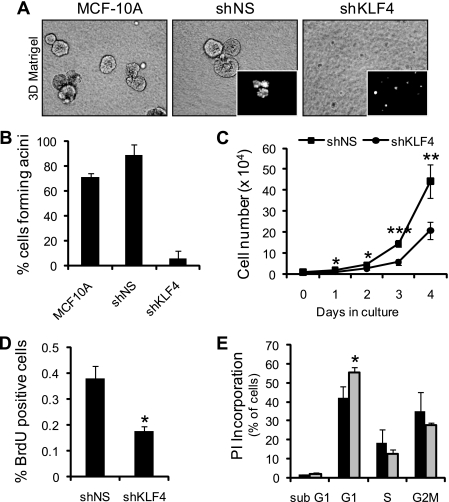
The Krüppel-like factor 4 (KLF4) is a transcriptional regulator of proliferation and differentiation in epithelial cells, both during development and tumorigenesis. Although KLF4 functions as a tumor suppressor in several tissues, including the colon, the role of KLF4 in breast cancer is less clear. Here, we show that KLF4 is necessary for maintenance of the epithelial phenotype in non-transformed MCF-10A mammary epithelial cells. KLF4 silencing led to alterations in epithelial cell morphology and migration, indicative of an epithelial-to-mesenchymal transition. Consistent with these changes, decreased levels of KLF4 also resulted in the loss of E-cadherin protein and mRNA. Promoter/reporter analyses revealed decreased E-cadherin promoter activity with KLF4 silencing, while chromatin immunoprecipitation identified endogenous KLF4 binding to the GC-rich/E-box region of this promoter. Furthermore, forced expression of KLF4 in the highly metastatic MDA-MB-231 breast tumor cell line was sufficient to restore E-cadherin expression and suppress migration and invasion. These findings identify E-cadherin as a novel transcriptional target of KLF4. The clear requirement for KLF4 to maintain E-cadherin expression and prevent epithelial-to-mesenchymal transition in mammary epithelial cells supports a metastasis suppressive role for KLF4 in breast cancer.


Figures

FIGURE 1.
FIGURE 1.
KLF4 is required for the maintenance of mammary epithelial cell morphology. A, immunoblot analysis of KLF4 and β-actin in MCF-10A cells stably transduced with either nonspecific shRNA control (shNS and MirNS) or shRNA against KLF4 (shKLF4 and MirKLF4). Cells were cultured in complete growth medium, ±10% serum. B, quantitative RT-PCR analysis of KLF4 mRNA in shNS control and shKLF4 cells. Relative KLF4 mRNA levels were normalized to the human TATA binding protein (TBP). C, mesenchymal morphology induced by loss of KLF4 in MCF-10A cells. Cells were plated at low (top row) and high (bottom row) densities. Cells were monitored for morphological changes using phase-contrast microscopy. D, increased migration of shKLF4 and MirKLF4 cells compared with shNS and MirNS controls. A total of 1 × 10⁵ cells was suspended in 100 μl of complete medium, seeded on Transwell migration inserts, and allowed to migrate toward complete medium for 20 h. Five fields per insert were counted. B and D, averages and standard deviations from three independent experiments done in triplicate. *, p < 0.02; **, p < 0.002; and ***, p < 0.0001.
FIGURE 2.
FIGURE 2.
KLF4 silencing results in loss of acinus formation and decreased proliferation of mammary epithelial cells. A, for three-dimensional cultures, 2.5 × 10⁵ cells were plated using the overlay method (47). Photomicrographs were taken after 10 days in culture. Insets are fluorescent images of the control and shKLF4 cells, which also express GFP. B, quantitation of mammary acini formation. By day 4, the majority of the cells in shNS control cultures had formed visible clusters of at least 8 cells, whereas <10% of shKLF4 cells formed clusters surpassing 4 cells in size. Error bars represent the standard deviations of two experiments done in duplicate. C, growth rate determination of shNS and shKLF4 cells. 1 × 10⁴ cells were plated in complete medium. At 24-h time intervals, cells were trypsinized and counted. D, BrdUrd incorporation of shNS and shKLF4 cells. Cells were plated at 50% confluency and allowed to grow for 72 h prior to incubation with BrdUrd. Slides were processed for immunofluorescence and scored for % BrdUrd positivity. E, cell cycle analysis of shNS (black bars) and shKLF4 (gray bars) cells using propidium iodide uptake and flow cytometry. Cells were plated at 50% confluency and allowed to grow for 48 h before being analyzed. Bars in C–E represent the averages and standard deviations from three independent experiments performed in triplicate. *, p < 0.05; **, p < 0.005; and ***, p < 5.0 × 10⁻⁵.
FIGURE 3.
FIGURE 3.
KLF4 is required to sustain E-cadherin expression in non-transformed mammary epithelial cells. A, Western blot analysis of E-cadherin, N-cadherin, p120, β-catenin, KLF4, and β-actin and B, quantitative RT-PCR analysis of E-cadherin mRNA in shNS and MirNS control versus shKLF4 and MirKLF4 cells. The graph represents the average -fold change and standard deviation from three independent experiments. *, p < 0.02; **, p < 1.0 × 10⁻⁷. C, immunofluorescence staining for E-cadherin (green) in shNS control and shKLF4 knockdown cells. Nuclear DNA is counterstained with 4′,6-diamidino-2-phenylindole (blue).
FIGURE 4.
FIGURE 4.
KLF4 binds and activates the E-cadherin promoter. A, schematic representation of the (−359/+30) proximal E-cadherin luciferase construct (Ecad-Luc). Black boxes (KLF4) represent GC-boxes containing putative KLF4 target sites. Numbered boxes depict two E-boxes located near the transcriptional start site. Gray arrows depict the location of the forward and reverse primers used for ChIP PCR amplification. B, E-cadherin promoter activity is lost in shKLF4 cells. The Ecad-Luc reporter plasmid or pGL3basic was cotransfected with a Renilla-expressing control (phRG-TK) into MCF-10A shNS control and shKLF4 cells. For KLF4 overexpression, parental MCF-10A cells were transfected 24 h prior to transduction with either AdGFP control or AdKLF4. Luciferase values, normalized to Renilla levels, were expressed as -fold change over shNS or AdGFP controls. Error bars represent standard deviations of three independent experiments performed in triplicate (*, p < 1.0 × 10⁻⁴; **, p < 1.0 × 10⁻⁵). C, ChIP analysis of KLF4 at the E-cadherin promoter. A KLF4 antibody or rIgG serum was used to immunoprecipitate DNA-protein complexes from MCF-10A cells. Binding of KLF4 at the E-cadherin promoter was enriched over rIgG control. Representative amplification of PCR products, using the primers described in A is shown. A molecular weight ladder is shown in the far right lane. Independent ChIP experiments were performed at least two times.
FIGURE 5.
FIGURE 5.
KLF4 induces expression of E-cadherin protein and a transition to epithelial morphology in the mesenchymal-like MDA-MB-231 breast cancer cells. A, Western blot analysis comparing KLF4 and E-cadherin levels in MCF-10A cells and MDA-MB-231 cells. B, Western blot of E-cadherin, KLF4, Krt18, and β-actin in MDA-MB-231 cells 72 h post transduction with empty vector control (AdGFP) or FLAG/HA-tagged KLF4 (AdKLF4) adenovirus. C, phase-contrast images of MDA-MB-231 cells transduced with AdGFP or AdKLF4 adenovirus. Images were captured 24 h post transduction.
FIGURE 6.
FIGURE 6.
KLF4 transcriptional activation of E-cadherin results in decreased migration and invasion of MDA-MB-231 cells. A, quantitative RT-PCR of KLF4 and E-cadherin mRNA levels in MDA-MB-231 cells transduced with either AdGFP control or AdKLF4 adenovirus. B, activation of the Ecad-Luc reporter in MDA-MB-231 cells overexpressing KLF4. The (−359/+30) Ecad-Luc reporter vector or pGL3 control was cotransfected with a Renilla-expressing control (phRG-TK) into MDA-MB-231 cells. Cells were transduced with either AdGFP control or AdKLF4 12 h later. Luciferase and Renilla activities were quantified 48 h post transfection. Luciferase values were normalized to Renilla levels and expressed as -fold change over AdGFP control cells. C, ChIP of KLF4 at the E-cadherin promoter in MDA-MB-231 cells. A FLAG antibody was used to immunoprecipitate DNA-protein complexes from both AdGFP and FLAG/HA-tagged AdKLF4 transduced MDA-MB-231 cells. Chromatin fragments were PCR-amplified with the same E-cadherin promoter primers described in Fig. 4A. AdGFP cells served as a negative control. Migration (D) and invasion (E) of AdGFP and AdKLF4/MDA-MB-231 cells. Cells were transduced with AdGFP or AdKLF4 48 h prior to trypsinization and plating onto Transwell supports for migration, or Matrigel-coated supports for invasion. Cells were allowed to migrate or invade through supports for 6 or 24 h, respectively. For A and C, cells were either harvested for RNA or fixed for ChIP analysis at 72 h post-transduction. A is a representative of three independent experiments performed in triplicate. Error bars represent the standard deviations. For B, error bars represent the standard deviations of three independent experiments performed in triplicate. For D and E, five fields were counted per insert. Data represent the average -fold change and standard deviations from three independent experiments performed in triplicate. *, p < 0.005; **, p < 5.0 × 10⁻⁷.

Similar articles

Cited by

References

    1. Shields J. M., Christy R. J., Yang V. W. (1996) J. Biol. Chem. 271, 20009–20017 - PMC - PubMed
    1. Chen X., Johns D. C., Geiman D. E., Marban E., Dang D. T., Hamlin G., Sun R., Yang V. W. (2001) J. Biol. Chem. 276, 30423–30428 - PMC - PubMed
    1. Segre J. A., Bauer C., Fuchs E. (1999) Nat. Genet. 22, 356–360 - PubMed
    1. Katz J. P., Perreault N., Goldstein B. G., Lee C. S., Labosky P. A., Yang V. W., Kaestner K. H. (2002) Development 129, 2619–2628 - PMC - PubMed
    1. Swamynathan S. K., Katz J. P., Kaestner K. H., Ashery-Padan R., Crawford M. A., Piatigorsky J. (2007) Mol. Cell. Biol. 27, 182–194 - PMC - PubMed

Publication types