Review
. 2010 Jun 6;7 Suppl 3(Suppl 3):S307-20.
doi: 10.1098/rsif.2010.0095.focus. Epub 2010 Mar 31.

Mechanosensitivity of ion channels based on protein-lipid interactions

Affiliations
Review

Mechanosensitivity of ion channels based on protein-lipid interactions

Kenjiro Yoshimura et al. J R Soc Interface. .

Abstract

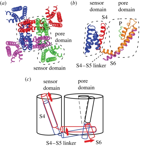
Ion channels form a group of membrane proteins that pass ions through a pore beyond the energy barrier of the lipid bilayer. The structure of the transmembrane segment of membrane proteins is influenced by the charges and the hydrophobicity of the surrounding lipids and the pressure on its surface. A mechanosensitive channel is specifically designed to change its conformation in response to changes in the membrane pressure (tension). However, mechanosensitive channels are not the only group that is sensitive to the physical environment of the membrane: voltage-gated channels are also amenable to the lipid environment. In this article, we review the structure and gating mechanisms of the mechanosensitive channels and voltage-gated channels and discuss how their functions are affected by the physical properties of the lipid bilayer.


Figures

Figure 1.
Figure 1.
Membrane protein embedded in lipid bilayer. (a) Cylindrical membrane protein in lipid bilayer when the height of the hydrophobic surface (grey) matches the thickness of hydrophobic core of the lipid bilayer (left). In a thinner bilayer, the hydrophobic mismatch is avoided either by rearrangement of the lipid molecules (middle) or by thinning of the protein (right). (b) The lateral pressure acting on the surface of the membrane protein (left). When the membrane protein is solubilized with detergent, the loss of the lateral pressure is likely to deform the transmembrane segment of the membrane protein (right).
Figure 2.
Figure 2.
Activation of bacterial mechanosensitive channels in a patch clamp experiment. When negative pressure (lower trace) is applied to the membrane in the patch clamp pipette (inset), mechanosensitive channel MscS is activated at low pressure. Further increase in pressure opens MscL. The amplitudes of single MscS and MscL channels are approximately 20 pA and approximately 75 pA, respectively.
Figure 3.
Figure 3.
Structure and function of the MscL mechanosensitive channel. (a) Crystal structure of M. tuberculosis MscL (PDB accession number: 2OAR). Each subunit is in a different colour. (b) Structure of single subunit of TbMscL. Shown are N-terminal helix (S1), the first and second transmembrane helices (TM1 and TM2), periplasmic loop (PL) and cytoplasmic helix (CP). (c) Positions of important residues in the schematic EcMscL. The pore is constricted between A20 and G26. TM2 is immersed in lipid bilayer between Y75 and K97. Gating threshold changes with the hydrophilicity of G22 and mechanosensitivity is lost on hydrophilic substitution of Y78. Charged residues RKK are involved in pH sensitivity and oligomer assembly. (d) Hydrophobic residues that hamper mechanosensiticity on asparagine substitution. Residues corresponding to EcMscL are indicated in orange in the space fill model of TbMscL. (e) Gating model of MscL. The closed structure is stabilized by the hydrophobic lock of the gate (i). The membrane tension perceived by the tension sensor opens the gate, which results in the exposure of the hydrophobic lock to water (ii). On full opening, the cytoplasmic helices are disassembled and the hydrophobic lock is buried in the protein interior by the rotation of TM1 (iii).
Figure 4.
Figure 4.
Structure and function of the MscS mechanosensitive channel. (a) Crystal structure of E. coli MscS (PDB accession number: 2OAU). Each subunit is in a different colour. (b) Structure of single subunit of MscS. Indicated are the first and second transmembrane helices (TM1 and TM2) and the third transmembrane helix, which is separated into two by a kink at G113 (TM3a and TM3b). (c) Important residues for the function of MscS. L105 and L109 form the constriction of the pore. D62 forms a salt bridge with R128 and R131. A kink at G113 occurs in the inactive state. (d) The gating threshold increases on asparagine substitution at the residues indicated by red (A34, I37, A85, L86), blue (I48, A51, L55), and orange (F68). Conversely, the threshold decreases on mutation at I39, V40 and I43 (green). Data from Nomura et al. (2006) and Okada et al. (2002).
Figure 5.
Figure 5.
Structure and conformational change of the potassium channel. (a) Crystal structure of Kv1.2 potassium channel (PDB accession number: 2A79). Each subunit is in a different colour. The pore domain is surrounded by four sensor domains. (b) Structure of a single subunit showing the position of S4 (red), selectivity filter (P), and gate-forming S6 (magenta). (c) Schematic representation of the conformational change that occurs on gating. Blue and red represent the position in the resting and activated state, respectively.

References

    1. Aggarwal S. K., MacKinnon R. 1996. Contribution of the S4 segment to gating charge in the Shaker K+ channel. Neuron 16, 1169–1177. (10.1016/S0896-6273(00)80143-9) - DOI - PubMed
    1. Ajouz B., Berrier C., Garrigues A., Besnard M., Ghazi A. 1998. Release of thioredoxin via the mechanosensitive channel MscL during osmotic downshock of Escherichia coli cells. J. Biol. Chem. 273, 26 670–26 674. (10.1074/jbc.273.41.26670) - DOI - PubMed
    1. Ajouz B., Berrier C., Besnard M., Martinac B., Ghazi A. 2000. Contributions of the different extramembranous domains of the mechanosensitive ion channel MscL to its response to membrane tension. J. Biol. Chem. 275, 1015–1022. (10.1074/jbc.275.2.1015) - DOI - PubMed
    1. Akitake B., Anishkin A., Liu N., Sukharev S. 2007. Straightening and sequential buckling of the pore-lining helices define the gating cycle of MscS. Nat. Struct. Mol. Biol. 14, 1141–1149. (10.1038/nsmb1341) - DOI - PubMed
    1. Anishkin A., Sukharev S. 2004. Water dynamics and dewetting transitions in the small mechanosensitive channel MscS. Biophys. J. 86, 2883–2895. (10.1016/S0006-3495(04)74340-4) - DOI - PMC - PubMed

Publication types

MeSH terms