. 2010 Sep 6;7(50):1293-9.
doi: 10.1098/rsif.2010.0096. Epub 2010 Mar 31.

Laminar flow around corners triggers the formation of biofilm streamers

Affiliations

Laminar flow around corners triggers the formation of biofilm streamers

Roberto Rusconi et al. J R Soc Interface. .

Abstract

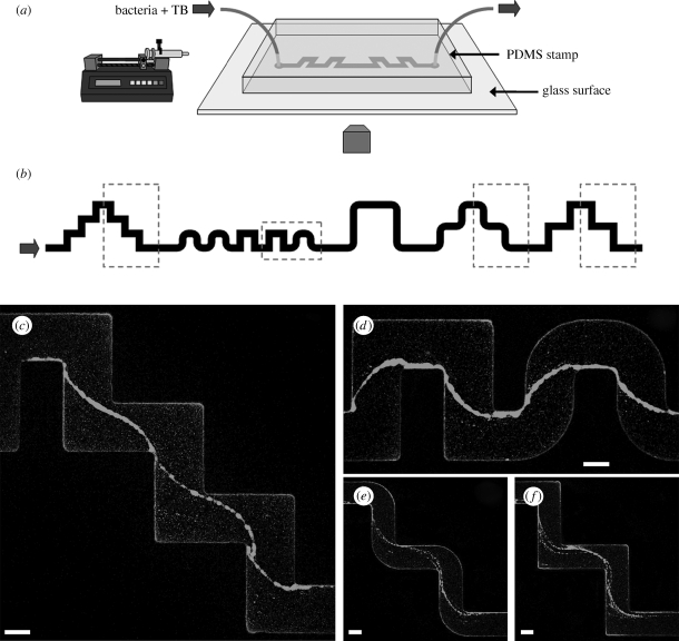
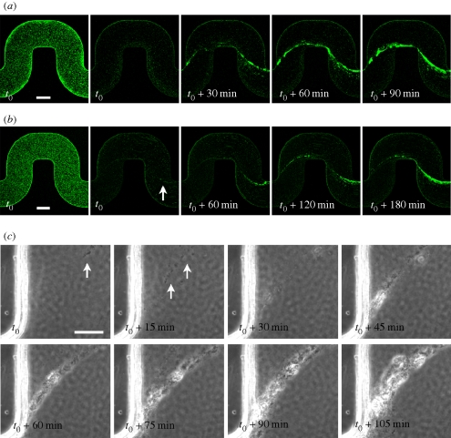
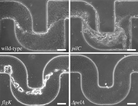
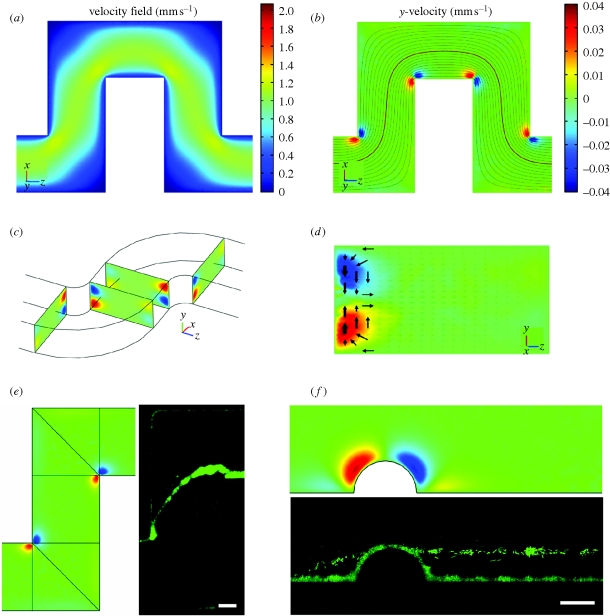
Bacterial biofilms have an enormous impact on medicine, industry and ecology. These microbial communities are generally considered to adhere to surfaces or interfaces. Nevertheless, suspended filamentous biofilms, or streamers, are frequently observed in natural ecosystems where they play crucial roles by enhancing transport of nutrients and retention of suspended particles. Recent studies in streamside flumes and laboratory flow cells have hypothesized a link with a turbulent flow environment. However, the coupling between the hydrodynamics and complex biofilm structures remains poorly understood. Here, we report the formation of biofilm streamers suspended in the middle plane of curved microchannels under conditions of laminar flow. Experiments with different mutant strains allow us to identify a link between the accumulation of extracellular matrix and the development of these structures. Numerical simulations of the flow in curved channels highlight the presence of a secondary vortical motion in the proximity of the corners, which suggests an underlying hydrodynamic mechanism responsible for the formation of the streamers. Our findings should be relevant to the design of all liquid-carrying systems where biofilms are potentially present and provide new insights on the origins of microbial streamers in natural and industrial environments.


Figures

Figure 1.
Figure 1.
(a) Schematic of the microfluidic set-up. (b) Layout of a portion of the channel used in the experiments. The width is 200 μm and the typical height is 100 μm. The round corners have an inner radius of curvature of 50 μm and an outer of 250 μm. The turns are spaced differently all along the channel by 400 μm (c,d), 600 μm (e,f) and 1200 μm. The direction of the flow is from left to right. (c,d) Streamers with wild-type GFP-labelled PA14 (OD600 = 0.25) after 12 h of constant flow at 0.75 μl min−1. (e,f) Streamers with wild-type GFP-labelled PA14 (OD600 = 0.4) after 10 h of constant flow at 0.75 μl min−1. The images are taken in the middle horizontal plane of the channel with a confocal microscope (scale bars, 100 μm).
Figure 2.
Figure 2.
(ad) Three-dimensional reconstruction of two streamers from confocal z-scan images. Above perspective (a,c) and horizontal views (b,d) are shown.
Figure 3.
Figure 3.
(a,b) Time evolution of the streamers: the first image in the sequences is taken at the bottom of the channel, the next four at the middle-height plane. The optical densities are (a) OD600 = 0.4 and (b) OD600 = 0.17. The time t0 corresponds to 6 h (a) and 7 h (b) of constant flow at 0.75 μl min−1. The white arrow indicates a very thin initial streamer. (c) Time sequence of phase-contrast microscopy images acquired every 1 min at 40× magnification (the focus is roughly in the middle of the channel). Time t0 corresponds to 5 h and 45 min of constant flow at 1 μl min−1. The initial concentration of PA14 is equivalent to OD600 = 0.5. White arrows point to single cells or isolated small clusters that are not moving with the mainstream flow but, instead, seem to be attached to very thin filaments of extracellular matrix (not visible; scale bars, (a,b) 100 μm; (c) 25 μm).
Figure 4.
Figure 4.
Phase-contrast images (the focus is approximately in the middle of the channel) after 12 h of continuous flow at 1 μl min−1. Comparison between wild-type, type IV pili defective mutant (pilC), flagellar-mediated motility defective mutant (flgK) and mutant (ΔpelA) defective in the synthesis of one of the main components of the extracellular matrix in P. aeruginosa PA14 biofilms (scale bars, 100 μm).
Figure 5.
Figure 5.
(a) Numerical results of the modulus of the velocity field in a plane at ¼ of the channel height from the upper surface. Coordinate system (x, y, z) and colour scale are shown. (b) Flow components perpendicular to the primary flow (represented here with streamlines) in the same plane as (a). Red and blue colours indicate here velocity components orthogonal to the plane of the channel, and directing, respectively, upwards (positive y) and downwards (negative y). (c) Prospective view of the channel showing pairs of counter-rotating vertices in cross-sectional planes right before and after the turns. (d) A cross-section of the channel, right after a turn. Arrows show the motion of fluid elements in the secondary flow. (e,f) Comparisons between numerical simulations of the secondary flow and experimental observations of the streamers, in a curved channel (e) and in a straight channel with a lateral hemi-cylindrical bump (f) (scale bars, (e) 50 μm; (f) 25 μm).

Similar articles

Cited by

References

    1. Balsa T. F. 1998. Secondary flow in a Hele-Shaw cell. J. Fluid Mech. 372, 25–44. (10.1017/S0022112098002171) - DOI
    1. Battin T. J., Kaplan L. A., Newbold J. D., Hansen C. M. E. 2003. Contributions of microbial biofilms to ecosystem processes in stream mesocosms. Nature 426, 439–442. (10.1038/nature02152) - DOI - PubMed
    1. Besemer K., Hödl I., Singer G., Battin T. J. 2009. Architectural differentiation reflects bacterial community structure in stream biofilms. ISME J. 3, 1318–1324 (10.1038/ismej.2009.73) - DOI - PubMed
    1. Brooks J. D., Flint S. H. 2008. Biofilms in the food industry: problems and potential solutions. Int. J. Food Sci. Tech. 43, 2163–2176. (10.1111/j.1365-2621.2008.01839) - DOI
    1. Costerton J. W., Lewandowski Z., Caldwell D. E., Korber D. R., Lappin-Scott H. M. 1995. Microbial biofilms. Annu. Rev. Microbiol. 49, 711–745. (10.1146/annurev.mi.49.100195.003431) - DOI - PubMed

Publication types

LinkOut - more resources