. 2010 May 15;21(10):1753-62.
doi: 10.1091/mbc.e09-12-1079. Epub 2010 Mar 31.

A domesticated piggyBac transposase plays key roles in heterochromatin dynamics and DNA cleavage during programmed DNA deletion in Tetrahymena thermophila

Affiliations

A domesticated piggyBac transposase plays key roles in heterochromatin dynamics and DNA cleavage during programmed DNA deletion in Tetrahymena thermophila

Chao-Yin Cheng et al. Mol Biol Cell. .

Abstract

Transposons comprise large fractions of eukaryotic genomes and provide genetic reservoirs for the evolution of new cellular functions. We identified TPB2, a homolog of the piggyBac transposase gene that is required for programmed DNA deletion in Tetrahymena. TPB2 was expressed exclusively during the time of DNA excision, and its encoded protein Tpb2p was localized in DNA elimination heterochromatin structures. Notably, silencing of TPB2 by RNAi disrupts the final assembly of these heterochromatin structures and prevents DNA deletion to occur. In vitro studies revealed that Tpb2p is an endonuclease that produces double-strand breaks with four-base 5' protruding ends, similar to the ends generated during DNA deletion. These findings suggest that Tpb2p plays a key role in the assembly of specialized DNA elimination chromatin architectures and is likely responsible for the DNA cleavage step of programmed DNA deletion.


Figures
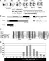
Figure 1.
Figure 1.
Characterization of TPB. (A) Tetrahymena nuclear developmental process during conjugation. (B) Schematic representation of Tpb proteins. (C) Comparison of the catalytic DDD-motif of Tpb2p and other piggyBac-like proteins. The DDD-motif of T. ni piggyBac consisting of D268, D346, and D447 is indicated. (D) Expression of TPB2 by quantitative RT-PCR. Total RNA extracted from vegetative (V), starved (S), and conjugating cells (2, 4, 6, 8, 10, 12, 14, 16, 18, and 20 h after postmixing) were used as templates. Quantification was normalized with α-tubulin mRNA expression.
Figure 2.
Figure 2.
TPB2 gene silence by hairpin RNA expression. (A) Schematic map of hairpin RNA constructs. Open arrows represent TPB2 gene with double lines inside indicating the region targeted by the hairpin RNA. (B) TPB2 gene silencing by hairpin RNA expression. Northern blot of total RNA samples extracted from conjugating cells (4, 6, 8, 10, 12, 14, and 16 h after postmixing) transformed with the hairpin RNA construct with or without inducing with 0.05 μg/ml CdCl2 in 10 mM Tris buffer. The approximate size of TPB2 mRNA is indicated to the left of the panels, and the right arrow indicates a new band likely from the degraded RNA. The robust TPB2 hairpin RNA expression was also detected. The RPL21 mRNA was used as an internal control to quantify relative folds of the full-length TPB2 mRNA level.
Figure 3.
Figure 3.
Phenotype of TPB2 silencing in conjugation. (A) Late conjugating stages analysis of the mutant. Conjugating cells (14, 20, and 30 h after postmixing) of TPB2 hairpin RNA-containing strains with or without induction with 0.05 μg/ml CdCl2 were stained with DAPI to reveal their nuclear stages (Figure 1A). NM (New Mac)-1, anlagen swelled in mating cells; NM-2, mating cells separated and two macronuclei and two micronuclei were present; NM-3, two macronuclei and one micronucleus were present. (B) Viability of progeny. Wild-type and TPB2 RNA hairpin cells were mated as indicated. Individual cell mating pairs were cloned into a drop of growth media at 10 h after postmixing. Cells were examined for progeny production by viability and drug resistance, which is specific for new developing macronucleus in progeny.
Figure 4.
Figure 4.
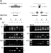
DNA elimination and chromosome breakage of TPB2 silencing in conjugation. (A) Schematic drawing of the IES elimination and chromosome breakage PCR assay. The open box represented DNA region retained in the macronucleus, and the filled box represented IES, Cbs, or telomere regions. Arrows indicate the positions of PCR primers. The relative lengths of the expected products were shown at top (Mic form) and bottom (Mac form). (B) The result of IES elimination and chromosome breakage PCR assay. The genomic DNA were isolated from pools of conjugating cells (30 h after postmixing) of wild type (1) and TPB2 hairpin RNA-containing strains (2 and 3) with or without induction with 0.05 μg/ml CdCl2. The open arrows indicate Mic form PCR product, and the filled arrows indicate Mac form PCR product.
Figure 5.
Figure 5.
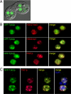
Localization of GFP-Tpb2p. (A) Overexpression of the N-terminal GFP-Tpb2p fusion protein. The GFP-Tpb2p fusion protein localized within the developing macronuclear anlagen in living conjugating cells. (B) Colocalization of GFP-Tpb2p and heterochromatin marker (H3K27me3, H3K9me3, and Pdd1p). Ten-hour conjugating cells with GFP-Tpb2p (green) were processed for immunofluorescence staining with the indicated antibodies (red). (C) Colocalization of GFP-Tpb2p and Pdd1p in the DNA elimination heterochromatin structure. Fourteen-hour conjugating cells expressing GFP-Tpb2p (green) were processed for immunofluorescence staining with an anti-Pdd1p antibody (red) and stained with DAPI for DNA (blue).
Figure 6.
Figure 6.
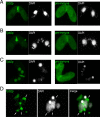
Localization of endogenous Tpb2p by antibody immunofluorescence staining. Cells were processed for immunofluorescence staining (green) and DAPI staining (white) using an anti-Tpb2p antibody and preimmune serum at different developmental stages in conjugation. (A) “Crescent” stage in micronuclei meiosis. (B) postmeiotic mitosis stage; (C) new developing macronuclei stage at early mating pair; (D) at the late conjugating stage after pair separation.
Figure 7.
Figure 7.
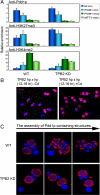
TPB2 is dispensable for recruiting heterochromatin markers to IES and is essential for the assembly of heterochromatin structures. (A) Conjugating cells of wild type (WT) and TPB2 knockdown strains (TPB2 KD) were processed for ChIP using the antibodies indicated. Two micronuclear IESs (M-mic and PGM1-mic) and two macronuclear genes (PGM1-mac and MTT1-mac) were assayed by quantitative PCR, and the relative enrichment values are shown. (B) Conjugating cells (12–16 h) of TPB2 hairpin RNA strains with or without induction with 0.05 μg/ml CdCl2 were processed for immunoflurescence staining with an anti-Pdd1p antibody (red) and DAPI staining (blue). (C) Magnified images of the assembly of Pdd1p-containing structures in WT and TPB2 KD cells.
Figure 8.
Figure 8.
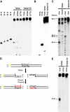
Tpb2p has DDD-motif dependent endonuclease activities. (A) Wild-type Tpb2p and a mutant Tpb2p in which all three aspartic acids in the DDD motif were replaced by leucines (Tpb2p-CD) were expressed in E. coli and incubated with 50-base pair DNA substrates that had TTAA, GTAG, or GTTG sequence once at their middle. One of two strands was 5′-end labeled. These products were separated in a denaturing polyacrylamide gel and detected by a phosphorimager. 23-, 24-, 25-, and 26-nt oligo DNAs were used as size markers. (B) The same product from the 50-base pair DNA substrate, which has TTAA shown in A, was separated in a native polyacrylamide gel and detected by a phosphorimager. Twenty-three- and 27-base pair oligo DNAs were used as size markers. (C) Tpb2p and Tpb2p-CD were expressed in E. coli and incubated with 50-base pair DNA substrates RArt or RArt30, which had AGTGAT sequence identified at a boundary of Tetrahymena R-element at their 25th and 30th position, respectively. One of two strands was 5′-end labeled. An aliquot of the product from RArt was also treated with Klenow fragment. These products were separated in a denaturing polyacrylamide gel and detected by a phosphorimager. Positions of 25-, 29-, 30-, and 50-nt oligo DNAs used as size markers are indicated in left. (D) Schematic representation of RArt cleavage by Tpb2p and followed by the fill-in reaction by Klenow fragment. 32P-labeled end is marked with yellow stars, and nucleotides added by Klenow fragment are highlighted by red. (E) Tpb2p and Tpb2p-CD were expressed in E. coli and incubated with 50-base pair DNA substrates which had TTAA or GTGA sequence once at their middle. One of two strands was 5′-end labeled. These products were separated in a denaturing polyacrylamide gel and detected by a phosphorimager. Twenty-three-nucleotide oligo DNA was used as size markers.

Similar articles

Cited by

References

    1. Agrawal A., Eastman Q. M., Schatz D. G. Transposition mediated by RAG1 and RAG2 and its implications for the evolution of the immune system. Nature. 1998;394:744–751. - PubMed
    1. Aronica L., Bednenko J., Noto T., DeSouza L. V., Siu K. W., Loidl J., Pearlman R. E., Gorovsky M. A., Mochizuki K. Study of an RNA helicase implicates small RNA-noncoding RNA interactions in programmed DNA elimination in Tetrahymena. Genes Dev. 2008;22:2228–2241. - PMC - PubMed
    1. Austerberry C. F., Allis C. D., Yao M. C. Specific DNA rearrangements in synchronously developing nuclei of Tetrahymena. Proc. Natl. Acad. Sci. USA. 1984;81:7383–7387. - PMC - PubMed
    1. Baudry C., Malinsky S., Restituito M., Kapusta A., Rosa S., Meyer E., Betermier M. PiggyMac, a domesticated piggyBac transposase involved in programmed genome rearrangements in the ciliate Paramecium tetraurelia. Genes Dev. 2009;23:2478–2483. - PMC - PubMed
    1. Borst P., Greaves D. R. Programmed gene rearrangements altering gene expression. Science. 1987;235:658–667. - PubMed

Publication types