. 2010 Jun 1;21(11):1788-98.
doi: 10.1091/mbc.e09-09-0774. Epub 2010 Mar 31.

RNF-121 is an endoplasmic reticulum-membrane E3 ubiquitin ligase involved in the regulation of beta-integrin

Affiliations

RNF-121 is an endoplasmic reticulum-membrane E3 ubiquitin ligase involved in the regulation of beta-integrin

Amir Darom et al. Mol Biol Cell. .

Abstract

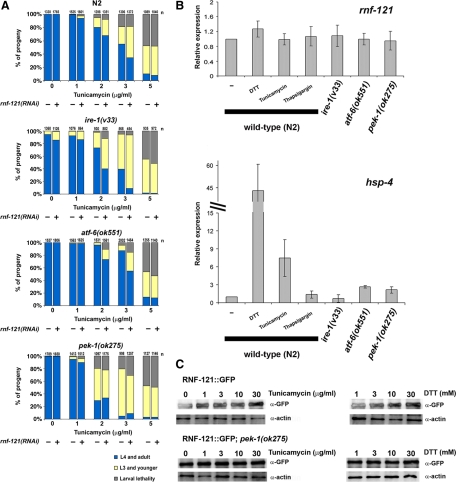
We report on the characterization of RNF-121, an evolutionarily conserved E3 ligase RING finger protein that is expressed in the endoplasmic reticulum (ER) of various cells and tissues in Caenorhabditis elegans. Inactivation of RNF-121 induced an elevation in BiP expression and increased the sensitivity of worms to ER stress. Genetic analysis placed RNF-121 downstream of the unfolded protein response (UPR) regulator protein kinase-like endoplasmic reticulum kinase (PERK). We identify PAT-3::GFP, the beta subunit of the heterodimeric integrin receptors, as an RNF-121 substrate; whereas induction of RNF-121 expression reduced the level of PAT-3::GFP in the gonad distal tip cells, inhibition of RNF-121 led to the accumulation of stably bound PAT-3::GFP inclusions. Correspondingly, overexpression of RNF-121 during early stages of gonad development led to aberrations in germline development and gonad migration that overlap with those observed after PAT-3 inactivation. The formation of these gonad abnormalities required functional ER-associated degradation (ERAD) machinery. Our findings identify RNF-121 as an ER-anchored ubiquitin ligase that plays a specific role in the ERAD pathway by linking it to the regulation of the cell adhesion integrin receptors.


Figures

Figure 1.
Figure 1.
Topology prediction, expression pattern, and in vitro E3 ligase activity of RNF-121. (A) Schematic representation of the predicted membrane topology of RNF-121. Six TMDs are predicted, the RING finger domain faces the cytosol. The amino acid sequence of the RING finger domain is shown. The cysteine and histidine residues of the RING finger domain are shown in red. The predicted E2 binding site (V224) is labeled in yellow (B and C) RNF-121::GFP is localized to the ER in body wall muscles (mu), hypodermis (hyp), and seam cells (SE). Bar, 10 μm. (D) RNF-121::GFP colocalizes with the mRFP::SP12 reporter. Bar, 5 μm. (E and F) RNF-121::GFP is expressed in the somatic gonad and vulva. Lateral view of midL4 animal. Expression is detected in the vulval cells (vul), spermathecal cells (sp), uterine cells (ut), and the distal tip cell (DTC). Bar, 10 μm. (G) RNF-121::GFP colocalizes with the mRFP::mans reporter. Bar, 5 μm. (H) In vitro self-ubiquitination assays were performed using wild-type GST-RNF-121RING or RING mutant forms of bacterially expressed and purified protein. In lane 4 (−), the reaction includes E1, E2, Ub, and ATP without the E3 ligase. GST-RNF-5 served as a positive control. Arrow indicates the E1 enzyme. Arrowhead indicates the 5% stacking gel-10% running gel boundary. High-molecular-weight smears generated by autoubiquitination of the indicated E3 are labeled with parenthesis. Coomassie-stained gel of the purified glutathione transferase (GST)-tagged expressed proteins is shown (bottom).
Figure 2.
Figure 2.
Inactivation of rnf-121 increases sensitivity to ER stress and induces the UPR. (A) N2 (wild-type) and rnf-121(ok848) embryos were treated with indicated concentrations of tunicamycin, and the various developmental stages were analyzed after 72-h incubation at 20°C. Animals were categorized into three groups: adults and L4, arrested L3 and younger and dead larvae. Each category is represented by the bar graph as the percentage of the total number of embryos (n = total number of embryos from 9 to 12 plates of 3 independent experiments, indicated above each bar). (B) Elevated basal expression of the hsp-4::gfp transcriptional reporter in rnf-121(RNAi)-treated worms. The hsp-4::gfp transgenic worms were fed with control (vector) or rnf-121(RNAi) bacteria in regular medium at 20°C. Arrow indicates the intestinal anterior cells.
Figure 3.
Figure 3.
RNF-121 is regulated by the PEK-1 pathway. (A) N2 (wild-type), ire-1(v33), pek-1(ok275), and atf-6(ok551) mutant worms were treated with control vector (−) or rnf-121(RNAi) (+) and were subjected to the tunicamycin sensitivity assay. The various developmental stages were analyzed after 72-h incubation at 20°C. Animals were categorized into three groups: adults and L4, arrested L3 and younger, and dead larvae. Each category is represented by the bar graph as the percentage of the total number of embryos ([n] indicated above each bar, total number of embryos from 9 to 12 plates of 3–6 independent experiments). (B) Differential expression of rnf-121 and hsp-4 after treatment with DTT, tunicamycin, and thapsigargin and in ire-1, atf-6, and pek-1 mutant worms. Relative quantification of each gene was determined using real-time RT-PCR. Mean values ± SEM from three independent treatments and RNA preparations are shown. Fold regulation was calculated relative to N2 (wild-type) nontreated animals. (C) RNF-121::GFP protein levels in wild-type (top) and pek-1(ok275) mutant worms (bottom) after treatment with tunicamycin and DTT. Total lysates (50 μg) were subjected to immunoblot analysis with anti-GFP and anti-actin antibodies.
Figure 4.
Figure 4.
Overexpression of RNF-121 at the L2 stage results in germline and gonad defects. (A and B) DIC and DAPI staining of wild-type adult hermaphrodite gonad arms. Marked are pachytene nuclei (arrowhead), diplotene (long arrow), and oocytes at diakinesis (short arrow). A scheme of the DTC path is shown on the left. (C–E) DIC and DAPI staining of hsp-16p::RNF-121 adult hermaphrodite gonad arms. Marked are pachytene nuclei (arrowhead) and oocytes at diakinesis (short arrow). The first oocyte is outlined. (F) Abnormal sperm in the spermatheca of hsp-16p::RNF-121 adult hermaphrodite (arrow). (G and H) DTCs migration defects in hsp-16p::RNF-121 adults. Bar, 10 μm. (I) Percentage of animals with abnormal gonads. Analysis was performed to the transgenic lines (3 independent lines from each construct) hsp-16p::RNF-121, hsp-16p::RNF-121C222AC225A, and hsp-16p::RNF-121 treated with der-1(RNAi). RNAi of der-1 in wild-type worms (N2 background) was performed as a control. Additional controls are N2 worms and transgenic worms expressing the E3 ligase RNF-5. Worms were treated with heat shock at mid-L2 stage to induce transgene expression (+) or nontreated controls (−). n = total number of worms analyzed in three to five independent experiments (indicated above each bar).
Figure 5.
Figure 5.
PAT-3::GFP degradation and accumulation depend on RNF-121 activity. (A) PAT-3::GFP expression in control (top) and in hsp-16p::RNF-121 (bottom). The nuclear envelope (NE) and surrounding reticulum of two body-wall muscle cells and the DTCs are shown in each panel (confocal images). Analysis was performed 3 h after heat shock at the mid-L2 stage. Muscle cell are outlined in red and DTCs in yellow. The NE is labeled with an arrowhead. Bar, 5 μm. (B) Distribution of the ratio of fluorescence intensities (DTC/muscle) in PAT-3::GFP (blue) and PAT-3::GFP; hsp-16p::RNF-121 (black) worms. Intensity values in the DTCs (yellow) are normalized to the intensity values in the nearest muscle nuclei (red) at the same focal plane. Analysis was performed on n = 50 worms from each strain (p < 0.0001) (C) In vitro ubiquitination of PAT-3::GFP immunoprecipitated from rnf-121(ok848); PAT-3::HA::GFP worms. Equal aliquots of immunopurified PAT-3::HA::GFP were used in each reaction in the presence of bacterially expressed and purified GST-RNF-121RING, E2, HA-ubiquitin and ATP, with or without E1 as indicated. Reactions were incubated at 37°C for 20 min and terminated with 8 M urea buffer. Black arrows indicate PAT-3::HA::GFP. Gray arrows indicate a putative spliced-form of PAT-3::HA::GFP. Arrowheads indicate the 5% stacking gel-8% running gel boundary. The ubiquitinated forms of PAT-3::HA::GFP are indicated by parentheses. IP, 25% of the immunoprecipitate used in each ubiquitination reaction. (D) PAT-3::GFP animals treated with control bacteria (empty L4440 vector; top) or rnf-121(RNAi) bacteria (bottom). Worms were grown in normal medium at 20°C. Variable sizes of fluorescent inclusions are shown (arrows) in PAT-3::GFP;rnf-121(RNAi) young adults. (E) Quantitative analysis of inclusion formation in PAT-3::GFP worms. Three independent experiments were performed (n = 150). Mean and SEM values are presented. Control worms were fed with empty L4440 vector. (F) Accumulation of fluorescent inclusions in rnf-121(ok848) worms (top) and immunoblot analysis of GFP and actin levels in total worm lysates (100 μg) from the indicated strains (bottom); rnf-121(ok848) worms were grown in liquid culture at 25°C (G) FRAP analysis of PAT-3::GFP inclusions in muscle cells of rnf-121(RNAi) animals. The boxed area inside the inclusion was photobleached. Example of a representative inclusion is shown (n > 15). (H) FLIP analysis of PAT-3::GFP inclusions in rnf-121(RNAi) animals. The marked region was repeatedly photobleached. Representative analysis is shown (n > 15). (I) PAT-3::GFP animals treated with 2 μg/ml tunicamycin accumulate smaller fluorescent inclusions. Bar, 5 μm.

References

    1. Amano T., et al. Synoviolin/Hrd1, an E3 ubiquitin ligase, as a novel pathogenic factor for arthropathy. Genes Dev. 2003;17:2436–2449. - PMC - PubMed
    1. Brenner S. The genetics of Caenorhabditis elegans. Genetics. 1974;77:71–94. - PMC - PubMed
    1. Broday L., Hauser C. A., Kolotuev I., Ronai Z. Muscle-epidermis interactions affect exoskeleton patterning in Caenorhabditis elegans. Dev. Dyn. 2007;236:3129–3136. - PubMed
    1. Broday L., Kolotuev I., Didier C., Bhoumik A., Podbilewicz B., Ronai Z. The LIM domain protein UNC-95 is required for the assembly of muscle attachment structures and is regulated by the RING finger protein RNF-5 in C. elegans. J. Cell Biol. 2004;165:857–867. - PMC - PubMed
    1. Brzovic P. S., Keeffe J. R., Nishikawa H., Miyamoto K., Fox D., 3rd, Fukuda M., Ohta T., Klevit R. Binding and recognition in the assembly of an active BRCA1/BARD1 ubiquitin-ligase complex. Proc. Natl. Acad. Sci. USA. 2003;100:5646–5651. - PMC - PubMed

Publication types

MeSH terms

LinkOut - more resources