. 2010 Jun;84(12):5848-59.
doi: 10.1128/JVI.00307-10. Epub 2010 Mar 31.

The Rab11 pathway is required for influenza A virus budding and filament formation

Affiliations

The Rab11 pathway is required for influenza A virus budding and filament formation

Emily A Bruce et al. J Virol. 2010 Jun.

Abstract

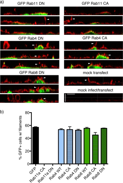
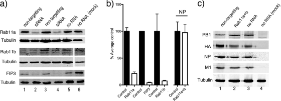
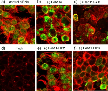
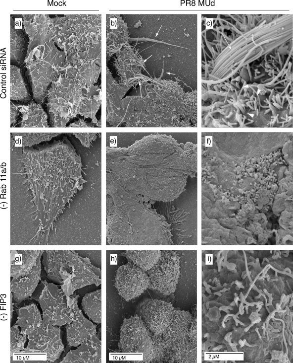
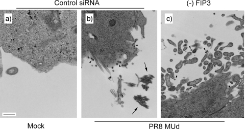
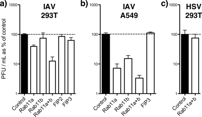
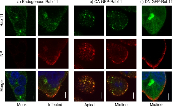
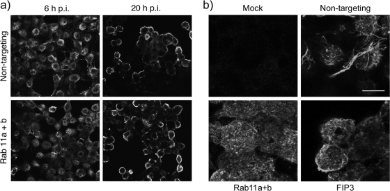
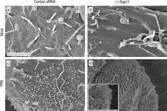
Influenza A virus buds through the apical plasma membrane, forming enveloped virus particles that can take the shape of pleomorphic spheres or vastly elongated filaments. For either type of virion, the factors responsible for separation of viral and cell membranes are not known. We find that cellular Rab11 (a small GTP-binding protein involved in endocytic recycling) and Rab11-family interacting protein 3 ([FIP3] which plays a role in membrane trafficking and regulation of actin dynamics) are both required to support the formation of filamentous virions, while Rab11 is additionally involved in the final budding step of spherical particles. Cells transfected with Rab11 GTP-cycling mutants or depleted of Rab11 or FIP3 content by small interfering RNA treatment lost the ability to form virus filaments. Depletion of Rab11 resulted in up to a 100-fold decrease in titer of spherical virus released from cells. Scanning electron microscopy of Rab11-depleted cells showed high densities of virus particles apparently stalled in the process of budding. Transmission electron microscopy of thin sections confirmed that Rab11 depletion resulted in significant numbers of abnormally formed virus particles that had failed to pinch off from the plasma membrane. Based on these findings, we see a clear role for a Rab11-mediated pathway in influenza virus morphogenesis and budding.


Figures

FIG. 1.
FIG. 1.
Effect of overexpressing GFP-tagged Rab polypeptides on influenza virus filament formation. CaCo2 cells were transfected (or mock transfected) with plasmids expressing the indicated GFP-tagged polypeptides and incubated overnight before infection at a multiplicity of infection of 10 with PR8 MUd. Cells were fixed at 16 h p.i., and cell surface HA (stained in red by anti-PR8 serum) and GFP (green) were imaged by confocal microscopy. (a) Representative z-plane reconstructions are shown. Scale bar, 5 μm. (b) A minimum of 200 cells from two independent experiments were scored for the presence or absence of filamentous projections. The mean and range are plotted.
FIG. 2.
FIG. 2.
Cellular and viral protein accumulation in siRNA-treated cells. 293T cells were transfected (or mock transfected) with siRNA sequences targeting the indicated cellular polypeptides. At 72 h post transfection the cells were infected (or mock infected where indicated) with PR8 virus at a multiplicity of infection of 5 for 24 h before further analysis. (a and c) Cells were lysed and analyzed by Western blotting for the indicated proteins. (b) Levels of the indicated proteins were quantified by densitometry, normalized to tubulin levels, and plotted as percentages of the average amount contained in cells treated with nontargeting control or no RNA. The means and standard deviations (Rab11a, n = 16; Rab 11b and FIP3, n = 4; NP, n = 4) are plotted. Note that the gel conditions used in the upper blot of panel c did not resolve PB1 and PB1-N40 (an N-terminally truncated variant of PB1) (56).
FIG. 3.
FIG. 3.
Influenza virus filament formation in siRNA-treated cells. 293T cells were transfected with siRNA sequences targeting the indicated proteins or with a nontargeting control (a and d). At 72 h posttransfection cells were infected (or mock infected) with PR8 MUd at a multiplicity of infection of 5, fixed at 16 h p.i., and stained with fluorescently tagged WGA to visualize the cell surface (red) as well as with anti-NP serum to validate infection (green). Images are maximum intensity projections of confocal z-stacks taken through the cell at approximately 0.5-μm steps.
FIG. 4.
FIG. 4.
SEM analysis of filament formation in Rab11- or FIP3-depleted (−) cells. 293T cells were transfected with siRNA sequences targeting Rab11a and Rab11b (d to f), FIP3 (g to i), or a nontargeting control (a to c), and infected with PR8 MUd at a multiplicity of infection of 5 at 72 h posttransfection. At 16 h p.i. cells were fixed and processed for SEM imaging.
FIG. 5.
FIG. 5.
TEM analysis of viral budding in FIP3-depleted cells. 293T cells were transfected with nontargeting siRNA sequences (a and b) or siRNA against FIP3 (c) and infected or mock infected as labeled with PR8 MUd at a multiplicity of infection of 5 at 72 h posttransfection. At 16 h p.i. cells were fixed and processed for TEM imaging. Scale bar, 500 nm. Arrowheads indicate spherical virus particles; arrows indicate cross sections through bundles of filaments.
FIG. 6.
FIG. 6.
Virus replication in siRNA-treated cells. 293T or A549 cells were transfected (or mock transfected) with siRNA sequences targeting the indicated cellular polypeptides. At 72 h posttransfection the cells were infected with PR8 virus (influenza A virus [IAV]) at a multiplicity of infection of 5 or with HSV at a multiplicity of infection of 10. Supernatants from infected cells were harvested at 24 h p.i., and titers were determined by plaque assay. The titers obtained from nontransfected cells and cells treated with nontargeting siRNAs were averaged and defined as 100%. The mean and standard error (IAV-infected 293T cells, n ≥4 replicate experiments; IAV-infected A549 cells, n ≥3; HSV-infected 293T cells, n = 2) are plotted.
FIG. 7.
FIG. 7.
Colocalization of Rab11 and influenza virus NP. (a) 293T cells were mock infected or infected as labeled with PR8 at a multiplicity of infection of 5, fixed at 6 h p.i., and stained for Rab11 (green), NP (red), and DNA (blue). (b and c) Cells were transfected with plasmids encoding GFP-tagged CA or DN Rab11 proteins, infected 18 h posttransfection, and stained at 6 h p.i. for NP and DNA. Single optical slices are shown. Scale bar, 4 μm.
FIG. 8.
FIG. 8.
NP and HA localization in Rab11- or FIP3-depleted cells. 293T cells were transfected with siRNA sequences targeting Rab11a and Rab11b, FIP3, or a nontargeting control as indicated and infected (or mock infected) with PR8 at a multiplicity of infection of 5 at 72 h post transfection. (a) At 6 and 20 h p.i. cells were fixed and permeabilized before staining for NP. Single optical slices are shown. Each panel represents a 150- by 135-μm window. (b) At 16 h p.i. cells were fixed and stained for HA. Maximum intensity projections of a set of optical sections through the upper halves of the cells are shown. Scale bar, 10 μm.
FIG. 9.
FIG. 9.
SEM visualization of virus budding in Rab11-depleted cells. 293T cells were transfected with siRNA sequences targeting Rab11a and Rab11b (b and d) or a nontargeting control (a and c), and either mock infected or infected with PR8 at a multiplicity of infection of 5 at 72 h posttransfection. At 16 h p.i. cells were washed and fixed overnight on coverslips before processing for SEM. Scale bar, 2 μm.
FIG. 10.
FIG. 10.
TEM visualization of budding virions in Rab11-depleted cells. 293T cells were transfected with siRNA sequences targeting Rab11a (b), Rab11a and Rab11b (d and f), or a nontargeting control (a, c, and e), and mock infected (e) or infected with PR8 at a multiplicity of infection of 5 at 72 h posttransfection. At 16 h p.i. cells were fixed and processed for TEM. Scale bar, 100 nm.

References

    1. Amorim, M. J., E. K. Read, R. M. Dalton, L. Medcalf, and P. Digard. 2007. Nuclear export of influenza A virus mRNAs requires ongoing RNA polymerase II activity. Traffic 8:1-11. - PubMed
    1. Benli, M., F. Doring, D. G. Robinson, X. Yang, and D. Gallwitz. 1996. Two GTPase isoforms, Ypt31p and Ypt32p, are essential for Golgi function in yeast. EMBO J. 15:6460-6475. - PMC - PubMed
    1. Bourmakina, S. V., and A. Garcia-Sastre. 2003. Reverse genetics studies on the filamentous morphology of influenza A virus. J. Gen. Virol. 84:517-527. - PubMed
    1. Brock, S. C., J. R. Goldenring, and J. E. Crowe, Jr. 2003. Apical recycling systems regulate directional budding of respiratory syncytial virus from polarized epithelial cells. Proc. Natl. Acad. Sci. U. S. A. 100:15143-15148. - PMC - PubMed
    1. Bruce, E. A., L. Medcalf, C. M. Crump, S. L. Noton, A. D. Stuart, H. M. Wise, D. Elton, K. Bowers, and P. Digard. 2009. Budding of filamentous and non-filamentous influenza A virus occurs via a VPS4 and VPS28-independent pathway. Virology 390:268-278. - PubMed

Publication types