. 2010 Jun;84(12):6103-18.
doi: 10.1128/JVI.02042-09. Epub 2010 Mar 31.

Interplay of RNA elements in the dengue virus 5' and 3' ends required for viral RNA replication

Affiliations

Interplay of RNA elements in the dengue virus 5' and 3' ends required for viral RNA replication

Peter Friebe et al. J Virol. 2010 Jun.

Abstract

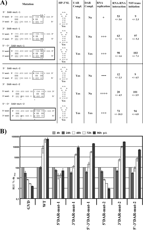
Dengue virus (DENV) is a member of the Flavivirus genus of positive-sense RNA viruses. DENV RNA replication requires cyclization of the viral genome mediated by two pairs of complementary sequences in the 5' and 3' ends, designated 5' and 3' cyclization sequences (5'-3' CS) and the 5' and 3' upstream of AUG region (5'-3' UAR). Here, we demonstrate that another stretch of six nucleotides in the 5' end is involved in DENV replication and possibly genome cyclization. This new sequence is located downstream of the AUG, designated the 5' downstream AUG region (5' DAR); the motif predicted to be complementary in the 3' end is termed the 3' DAR. In addition to the UAR, CS and DAR motifs, two other RNA elements are located at the 5' end of the viral RNA: the 5' stem-loop A (5' SLA) interacts with the viral RNA-dependent RNA polymerase and promotes RNA synthesis, and a stem-loop in the coding region named cHP is involved in translation start site selection as well as RNA replication. We analyzed the interplay of these 5' RNA elements in relation to RNA replication, and our data indicate that two separate functional units are formed; one consists of the SLA, and the other includes the UAR, DAR, cHP, and CS elements. The SLA must be located at the 5' end of the genome, whereas the position of the second unit is more flexible. We also show that the UAR, DAR, cHP, and CS must act in concert and therefore likely function together to form the tertiary RNA structure of the circularized DENV genome.


Figures

FIG. 1.
FIG. 1.
Characterization of DENV reporter replicon. (A) Schematic presentation of the replicons used in this study. pDRep has been described previously (8). In 5′ UTR-Cap-tr, the EMCV IRES is positioned downstream of the DENV 5′ end containing the first 72 nt of the capsid-coding region (C72nt), mediating translation of Renilla luciferase (Luciferase) reporter gene and the viral open reading frame (ORF) spanning the C-terminal sequence of E and NS1 to NS5. Luciferase is cleaved from the viral proteins by an engineered FMDV2A (FMDV) cleavage site. RNA structures in the 5′ and 3′ ends are indicated schematically. In 5′ UTR-Cap-tr-SPACER, the DENV 5′ end and the EMCV IRES are separated by an ∼650-nt-long spacer sequence derived from the GFP-coding sequence (SPACER). (B) Replication competence of DENV reporter replicons over a time course of 96 h posttransfection (p.t.). Time points are indicated on top, and RLU are expressed as a percentage of the value measured 4 h p.t., which was set to 100%. The replicons pDRep and GVD served as positive and negative controls, respectively. Results from at least three independent experiments are shown. Error bars reflect standard deviations. (C) Comparison of viral RNA copy number in BHK cells 96 h p.t. for the indicated replicon RNAs. RNA copy number was determined by qRT-PCR using NS5-specific primers and probe. The amount of viral RNA measured for the parental pDRep replicon was normalized to 18S RNA and set to 100%. All other normalized RNA levels are given as a percentage of pDRep. Results from at least three independent experiments are shown. Error bars reflect standard deviations.
FIG. 2.
FIG. 2.
Secondary structure of the DENV2 5′ end in the presence of the 3′ UTR and of the 3′ SL in the absence of 5′ RNA. (A) Interaction between the DENV 5′ and 3′ ends is depicted as determined from the solution structure of the 5′ UTR in the presence of the 3′ UTR (adapted from reference 25). Sequences derived from the DENV2 (strain 16681) 5′ end are on top, and those from the 3′ end are on the bottom. Structural elements are indicated (SLA, cHP), and known nucleotides required for 5′-3′ base pairing are boxed (5′-3′ UAR, 5′-3′ CS). The third double-stranded region between the 5′ and 3′ ends is surrounded by a dotted frame and magnified on top (DAR). Nucleotides involved in alternative stem formation at the 3′ end in the absence of 5′ RNA are underlined and shaded, and the structure itself is displayed below in 3′-5′ orientation (for details, refer to the legend to panel B). The start codon is highlighted in italics and marked with an asterisk. (B) Schematic presentation of the 3′ SL in the absence of 5′ RNA. The small stem-loop at the bottom of the 3′ SL is highlighted (dashed frame) and designated hairpin at 3′ SL (HP-3′ SL). Nucleotides involved in stem formation are underlined and shaded in all RNA structures (A and B).
FIG. 3.
FIG. 3.
The DAR sequences are important for RNA replication. (A) Overview of mutations and assay results. Detailed overview of mutations introduced into the 5′ UTR-Cap-tr-SPACER replicon are presented on the left side. The names of the mutants are indicated on top, with the other sequence as WT unless it is indicated that both the 5′ and 3′ mutant sequences are presented. Complementary nucleotides between 5′ and 3′ ends are indicated by a dash, and nucleotides involved in HP-3′ SL formation are underlined. The DAR motif is boxed. Mutations are highlighted with an asterisk. Mutants with restored complementarity between 5′-3′ DAR sequences are boxed. Prediction of HP-3′ SL formation is given in the column “HP-3′ SL” for each mutant. RNA replication levels are summarized with up to five plus signs, and no replication is shown with dashes; please refer to the legend to panel B for more details. Results regarding 5′-3′ RNA-RNA interaction and NS5 trans-initiation activity for each mutant are given as percentages of the quantified amount of 3′ RNA shifted (RNA-RNA interaction) or 3′ RNA synthesized compared to the 5′-3′ WT control. Values reflect mean values from at least three independent experiments, with the standard deviation (SD) shown immediately below. (B) Replication competence of variant DENV reporter replicons. RLU was measured over a time course of 96 h after transfection of variant replicons harboring the mutations indicated below the graph. For more details, refer to the legend to Fig. 1B. (C) RNA mobility shift analysis showing the effect of mutations within the 5′ and 3′ DAR and nearby sequences. Detailed overview of mutations are given in (A); mutant upstream DAR and downstream DAR contain the following 5′ sequence (limited to the sequence shown in panel A): 5′-CUGUACUUAUUCCAACGGAAA-3′ and 5′-CUGAUGAAUAACCAACGCUUU-3′; DAR sequences are underlined, mutations highlighted in bold). The 5′ RNA consists of the first 160 nt of the DENV genome, containing the mutations indicated along the top of the gel. The uniformly labeled 3′ SL RNA includes the final 106 nt of the DENV RNA genome, and the mutations are indicated below the gel. The ratio between the 5′ and 3′ RNAs was 100:1. The mobility of the 3′ SL alone or in complex with 5′ RNA is indicated on the left. The gel displayed is representative of results of at least three independent experiments. (D) Effects of mutations on RNA template usage and trans-initiation of RNA synthesis by purified RdRp. The gel displays the radiolabeled products from in vitro RdRp activity assays using the recombinant DENV NS5 RdRp domain. Mutations in the RNAs used as template (0.5 μg each) are indicated below (3′ UTR RNA template) and above (5′ UTR RNA template, corresponding to the first 160 nt) the gel. The migration pattern of the DENV 5′ UTR and 3′ UTR RNAs, as determined using the corresponding 5′ or 3′ in vitro-transcribed WT RNAs, are indicated on the left. Results displayed are representative of results of at least three independent experiments.
FIG. 4.
FIG. 4.
Primary sequences and complementarity of the DAR elements and the HP-3′ SL are involved in DENV RNA replication. (A) Overview of mutations analyzed and results obtained. Detailed overview of mutations introduced into the 5′ UTR-Cap-tr-SPACER replicon on the left side, with names indicated on top. Please refer to the legend to Fig. 3A for more details. Mutants with restored complementarity between 5′-3′ UAR and DAR sequences are boxed. (B) Replication levels of indicated mutant replicons over a time course of 96 h p.t. For more details, see the legend to Fig. 3B.
FIG. 5.
FIG. 5.
Loss of replication due to 5′ DAR mutation can be rescued by restored DAR complementarity. (A) Overview of mutations and assay results. Detailed overview of mutations introduced into the 5′ UTR-Cap-tr-SPACER replicon on the left side, with names indicated on top. Please refer to the legend to Fig. 3A for more details. Mutants with restored complementarity between 5′-3′ DAR sequences are boxed. (B) Detailed replication levels of indicated mutant replicons over a time course of 96 h p.t. For more details, see the legend to Fig. 3B.
FIG. 6.
FIG. 6.
Mutations introduced into the DENV 5′ end. The position of mutations within the 5′ end of the viral RNA are shown on top of the 5′-3′ RNA-RNA structure (see Fig. 2 for more details), indicated in bold and marked with an asterisk. The position of the mutations is further indicated by arrows, and the character of the mutations and their effects on RNA structure as predicted by mfold are shown, with mutated nucleotides marked with an asterisk. Names are indicated next to each mutation. Mutations were introduced only into the 5′ end. Mutated nucleotides are slightly enlarged. The start codon is shown in italics and underlined.
FIG. 7.
FIG. 7.
The SLA is 5′-position dependent. (A) Effects of mutations in the 5′ end on RNA replication in the context of the 5′UTR-Cap-tr-SPACER replicon. RLU was monitored over a time course of 96 h. The name and a schematic diagram of the 5′ end of the replicon is provided above the graph, indicating the region into which mutations were introduced. The names of the mutants are provided below the graph. The results are clustered into sections containing controls (Control), RNAs with mutations that destroy replication competence (non-viable), and RNAs with mutations resulting in viable replicons (viable). For more details, please refer to legend to Fig. 1B. (B) Rescue of mutations in the 5′ end by internal insertion of the 5′ UTR and capsid-coding region. A schematic diagram of the 5′ end of the replicon is provided above the graph, and the region into which mutations were introduced is indicated. The replication competence of the mutants identified below the graph is shown. As described above, the results were clustered into control, viable, and nonviable replicon sections. For more details, refer to the legend to panel A.
FIG. 8.
FIG. 8.
Two functional units are present in the viral 5′ end: the SLA and the UAR, DAR, cHP, and CS elements. (A) Rescue of mutations in the 5′ end by internal insertion of the 5′ UTR and capsid-coding region without the SLA. A schematic diagram of the 5′ end of the replicon upstream of the EMCV IRES is provided above the graph, and the region into which mutations were introduced is indicated. For a more-detailed description, refer to the legend to Fig. 7. (B) Analysis of the interplay between different RNA elements in the 5′ end. A “NoCS” mutation is present at the very 5′ end, and the mutations indicated below the graph were engineered into the inserted 5′ UTR-capsid-coding sequence fragment; as before, a schematic overview is provided on top. For more details, please refer to legend to Fig. 7.
FIG. 9.
FIG. 9.
Sequences outside the UAR, DAR and CS are not required for 5′-3′ RNA-RNA complex formation. RNA mobility shift analysis showing the effect of mutations within the DENV 5′ end on interaction with the 3′ end. The 5′ RNA consists of the first 160 nt of the DENV genome, carrying the mutations indicated on top of the gel. The uniformly labeled 3′ SL RNA includes the last 106 nt of the DENV genome. The ratio between the 5′ and 3′ RNAs was 100:1. The size of the 3′ SL alone or in complex with the 5′ DENV RNA is indicated on the left. The gel displayed here is representative of results of at least three independent experiments. For more details, refer to the legend to Fig. 3C.

Similar articles

Cited by

References

    1. Alvarez, D. E., C. V. Filomatori, and A. V. Gamarnik. 2008. Functional analysis of dengue virus cyclization sequences located at the 5′ and 3′UTRs. Virology 375:223-235. - PubMed
    1. Alvarez, D. E., A. L. Lella Ezcurra, S. Fucito, and A. V. Gamarnik. 2005. Role of RNA structures present at the 3′UTR of dengue virus on translation, RNA synthesis, and viral replication. Virology 339:200-212. - PubMed
    1. Alvarez, D. E., M. F. Lodeiro, C. V. Filomatori, S. Fucito, J. A. Mondotte, and A. V. Gamarnik. 2006. Structural and functional analysis of dengue virus RNA. Novartis Found. Symp. 277:120-132. - PubMed
    1. Alvarez, D. E., M. F. Lodeiro, S. J. Luduena, L. I. Pietrasanta, and A. V. Gamarnik. 2005. Long-range RNA-RNA interactions circularize the dengue virus genome. J. Virol. 79:6631-6643. - PMC - PubMed
    1. Astier-Gin, T., P. Bellecave, S. Litvak, and M. Ventura. 2005. Template requirements and binding of hepatitis C virus NS5B polymerase during in vitro RNA synthesis from the 3′-end of virus minus-strand RNA. FEBS J. 272:3872-3886. - PubMed

Publication types

LinkOut - more resources