. 2010 Jun;84(11):5656-69.
doi: 10.1128/JVI.00211-10. Epub 2010 Mar 31.

Murine coronavirus delays expression of a subset of interferon-stimulated genes

Affiliations

Murine coronavirus delays expression of a subset of interferon-stimulated genes

Kristine M Rose et al. J Virol. 2010 Jun.

Abstract

The importance of the type I interferon (IFN-I) system in limiting coronavirus replication and dissemination has been unequivocally demonstrated by rapid lethality following infection of mice lacking the alpha/beta IFN (IFN-alpha/beta) receptor with mouse hepatitis virus (MHV), a murine coronavirus. Interestingly, MHV has a cell-type-dependent ability to resist the antiviral effects of IFN-alpha/beta. In primary bone-marrow-derived macrophages and mouse embryonic fibroblasts, MHV replication was significantly reduced by the IFN-alpha/beta-induced antiviral state, whereas IFN treatment of cell lines (L2 and 293T) has only minor effects on replication (K. M. Rose and S. R. Weiss, Viruses 1:689-712, 2009). Replication of other RNA viruses, including Theiler's murine encephalitis virus (TMEV), vesicular stomatitis virus (VSV), Sindbis virus, Newcastle disease virus (NDV), and Sendai virus (SeV), was significantly inhibited in L2 cells treated with IFN-alpha/beta, and MHV had the ability to rescue only SeV replication. We present evidence that MHV infection can delay interferon-stimulated gene (ISG) induction mediated by both SeV and IFN-beta but only when MHV infection precedes SeV or IFN-beta exposure. Curiously, we observed no block in the well-defined IFN-beta signaling pathway that leads to STAT1-STAT2 phosphorylation and translocation to the nucleus in cultures infected with MHV. This observation suggests that MHV must inhibit an alternative IFN-induced pathway that is essential for early induction of ISGs. The ability of MHV to delay SeV-mediated ISG production may partially involve limiting the ability of IFN regulatory factor 3 (IRF-3) to function as a transcription factor. Transcription from an IRF-3-responsive promoter was partially inhibited by MHV; however, IRF-3 was transported to the nucleus and bound DNA in MHV-infected cells superinfected with SeV.


Figures

FIG. 1.
FIG. 1.
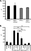
MHV rescues SeV from the antiviral effects of IFN when MHV infection is established before IFN treatment. (A) L2 fibroblasts cultures were mock treated or treated with 1,000 U/ml recombinant mouse IFN-α 3 h prior to SeV infection at an MOI of 1. At 16 h postinfection, SeV-infected cells were stained with SeV-specific monoclonal antibody and DAPI (4′,6-diamidino-2-phenylindole) to determine the level of infection. (B) L2 cells were infected with MHV (rA59/SMHV-2-EGFP) at an MOI of 1 for 3 h prior to treatment with 0 U/ml (bottom panels) or 1,000 U/ml (top and middle panels) recombinant mouse IFN-α. Following 3 h of IFN-α treatment, cultures were infected with SeV at an MOI of 1, fixed at 16 h postinfection, and subsequently stained with SeV-specific antibody. rA59/SMHV-2-EGFP infection was monitored by measuring levels of GFP expression. Data are representative of the results of four independent experiments.
FIG. 2.
FIG. 2.
MHV infection prior to IFN-α/β treatment inhibits activation of the ISRE. 293T cells were transiently transfected with expression vector for the MHV receptor CEACAM1a, an ISRE reporter construct linked to firefly luciferase (pElu-ISRE), and a renilla expression vector driven by the SV40 promoter (pRL-SV40). At 24 h posttransfection, the cultures were treated with 1,000 U/ml recombinant human IFN-α or IFN-β and simultaneously mock infected or infected with MHV at an MOI of 1 (A) or infected with MHV and treated with 1,000 U/ml IFN-α or -β 3 h later (B). Alternatively, cells were transfected with an expression vector for the V protein of simian virus 5 (V) for 24 h and then treated with 1,000 U/ml IFN-β. Cell lysates were collected 8 h posttreatment and assayed for firefly and renilla luciferase expression levels. Firefly luciferase values are normalized to renilla luciferase values. IFN-β-induced luciferase expression was set to 100%, and all other values were calculated as percentages of the IFN-β signal. Statistical significance was determined by Student's t test (**, P < 0.005). Data are representative of the results of four independent experiments.
FIG. 3.
FIG. 3.
MHV infection delays expression of a subset of ISGs induced by IFN-β. 293T cells were transfected with an expression vector for the MHV receptor. At 24 h later, cells were mock infected or infected with MHV at an MOI of 1. Cultures were treated with 0 U/ml (mock and MHV) or 1,000 U/ml recombinant human IFN-β 3 h post MHV or mock infection (MHVpre or mock pre). Total RNA was isolated from cells 8 h (A) or 15 h (B) post IFN-β treatment and analyzed for expression of indicated ISGs by performing qRT-PCR with gene-specific primers. Data are representative of the results of three independent experiments. Statistical significance was determined by Student's t test (*, P < 0.05; **, P < 0.005).
FIG. 4.
FIG. 4.
MHV does not alter STAT1 translocation to the nucleus or STAT1 and STAT2 phosphorylation. (A) An expression vector for STAT1 fused to GFP (STAT1-GFP) was transfected along with a plasmid expressing the MHV receptor into 293T cells. Cultures were then mock infected or infected with MHV at an MOI of 1 24 h posttransfection. MHV infection was allowed to proceed for 3 h before treatment of cultures with 0 U/ml (top panels) or 1,000 U/ml (middle and bottom panels) recombinant human IFN-β. Following 6 h of IFN-β treatment, cells were fixed in paraformaldehye and stained with an antibody that specifically detects MHV nucleocapsid protein. (B) 293T cells transiently transfected with MHV receptor were treated as described above, and cell lysates were collected at the indicated times post IFN-β treatment. Protein levels were normalized, and lysates were processed on sodium dodecyl sulfate-polyacrylamide gel electrophoresis (SDS-PAGE) gels for Western blot analysis with antibodies to detect STAT1 and STAT2 and phosphorylated forms of both proteins. Levels of α-tubulin were used to ensure equal levels of loading. Data are representative of the results of two independent experiments.
FIG. 5.
FIG. 5.
Preinfection of 293T with MHV delays ISG induction mediated directly by SeV. (A to C) 293T or Vero E6 cultures were transiently transfected with MHV receptor, pSV40-RL, and ISRE-luciferase reporter construct. (A) At 24 h later, cultures were left untreated (UT), infected with MHV (MHV), or mock infected (mock). Following MHV infection for 1 h (1h pre), 2 h (2h pre), or 3 h (3h pre), cultures were infected with SeV at an MOI of 1. Cell lysates were collected 8 h postinfection and analyzed for luciferase expression. Following 24 h transfection, 293T (B) or Vero E6 (C) cultures were left untreated, mock infected (mock pre), or infected with MHV (MHV pre). At 3 h post MHV infection, cultures were superinfected with SeV or a combination of MHV and SeV (MHV + SeV). Cells were lysed 8 h later and analyzed for firefly and renilla luciferase expression. Firefly luciferase levels were normalized to renilla luciferase levels. SeV-induced luciferase expression was set to 100%, and all other values were calculated as percentages of the SeV signal. Statistical significance was determined by Student's t test (*, P < 0.05; **, P < 0.005). Data represent the results of at least three independent experiments.
FIG. 6.
FIG. 6.
MHV delays induction of ISGs mediated by SeV. 293T cells were transfected with an expression vector for the MHV receptor. At 24 h later, cells were mock infected (mock pre) or infected with MHV (MHV pre) at an MOI of 1. Following 3 h of incubation with MHV, cultures were mock infected or infected with SeV (MOI = 1). Total RNA was isolated from cells 8 h (A) or 15 h (B) post SeV infection and analyzed for expression of indicated ISGs by performing qRT-PCR with gene-specific primers. The experiments were performed in triplicate, and statistical significance was determined by Student's t test (* P < 0.05; **, P < 0.005). Data represent the results of three independent experiments.
FIG. 7.
FIG. 7.
MHV reduces the ability of SeV to activate IRF-3 and NF-κB. 293T cells were transfected with expression plasmid for MHV receptor, pRL-SV40, and either of two IRF-3-responsive promoters, p55C1B (A) or pLuc-(PRDIII-I)3 (B and C), with or without pCAGGS-NS1. Alternatively, cells were transfected with pRL-SV40 and an NF-κB responsive promoter, pLuc-(PRDII)2 (D and E). At 24 h posttransfection, cultures were mock infected (mock pre) or infected with MHV (MHV pre) and then infected 3 h later with SeV (MOI = 1), treated with 1,000 U/ml TNF-α, or left untreated. Cells were lysed, and renilla expression levels were determined 8 h (A, B, and D) or 15 h (C and E) postinfection or posttreatment. (F) 293T cells were transiently transfected with pElu-ISRE and either pRL-SV40 or pRL-TK expressing renilla luciferase under the control of SV40 or thymidine kinase promoters, respectively. At 24 h posttransfection, cultures were left untreated (−) or infected with SeV or MHV and cell lysates were collected for analysis of luciferase expression 8 h postinfection. (A to E) Values represent percentages of firefly expression relative to SeV-treated culture values after normalization to renilla expression levels. Statistical significance was determined by Student's t test (*, P < 0.05; **, P < 0.005). Data represent the results of three independent experiments.
FIG. 8.
FIG. 8.
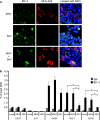
MHV does not prevent SeV-mediated IRF-3 translocation and DNA binding. 293T cells were transfected with an expression vector for the MHV receptor. (A) At 24 h posttransfection, cells were either left untreated or inoculated with MHV (top and bottom panels) at an MOI of 1. Following 3 h of incubation with MHV, cultures were mock infected (top panels) or infected with SeV (middle and bottom panels) at an MOI of 1. At 6 h post SeV infection, cells were fixed in paraformaldehyde and analyzed for immunofluorescence detection of IRF-3 and MHV nucleocapsid by the use of specific antibodies. (B) 293T cells seeded in 10-cm-diameter dishes were transfected with an expression vector for the MHV receptor and either empty control vector or pCAGGS-NS1. At 24 h posttransfection, cultures were either mock infected, infected with MHV (MOI = 1), or left untreated (NS1). At 3 h later, cells were inoculated with SeV (MOI = 1) and SeV infection was allowed to proceed for 6 h before cells were fixed for ChIP assays using an IRF-3-specific antibody or an IgG isotype control. qPCR analysis of precipitated DNA fragments by the use of primers designed within the promoter of specific genes was used to quantify IRF-3 binding to specific genes. Statistical significance was determined by Student's t test (*, P < 0.05; **, P < 0.005). Data represent the results of two independent experiments.

References

    1. Andersen, J., S. VanScoy, T. F. Cheng, D. Gomez, and N. C. Reich. 2008. IRF-3-dependent and augmented target genes during viral infection. Genes Immun. 9:168-175. - PubMed
    1. Andrejeva, J., D. F. Young, S. Goodbourn, and R. E. Randall. 2002. Degradation of STAT1 and STAT2 by the V proteins of simian virus 5 and human parainfluenza virus type 2, respectively: consequences for virus replication in the presence of alpha/beta and gamma interferons. J. Virol. 76:2159-2167. - PMC - PubMed
    1. Aurisicchio, L., P. Delmastro, V. Salucci, O. G. Paz, P. Rovere, G. Ciliberto, N. La Monica, and F. Palombo. 2000. Liver-specific alpha 2 interferon gene expression results in protection from induced hepatitis. J. Virol. 74:4816-4823. - PMC - PubMed
    1. Basler, C. F., A. Mikulasova, L. Martinez-Sobrido, J. Paragas, E. Muhlberger, M. Bray, H. D. Klenk, P. Palese, and A. Garcia-Sastre. 2003. The Ebola virus VP35 protein inhibits activation of interferon regulatory factor 3. J. Virol. 77:7945-7956. - PMC - PubMed
    1. Baum, A., and A. Garcia-Sastre. 31 October 2009, posting date. Induction of type I interferon by RNA viruses: cellular receptors and their substrates. Amino Acids. [Epub ahead of print.] doi:10.1007/s00726-009-0374-0. - DOI - PMC - PubMed

Publication types