. 2010 May;24(5):941-52.
doi: 10.1210/me.2009-0395. Epub 2010 Mar 31.

Cyclin-dependent kinase 5 modulates the transcriptional activity of the mineralocorticoid receptor and regulates expression of brain-derived neurotrophic factor

Affiliations

Cyclin-dependent kinase 5 modulates the transcriptional activity of the mineralocorticoid receptor and regulates expression of brain-derived neurotrophic factor

Tomoshige Kino et al. Mol Endocrinol. 2010 May.

Abstract

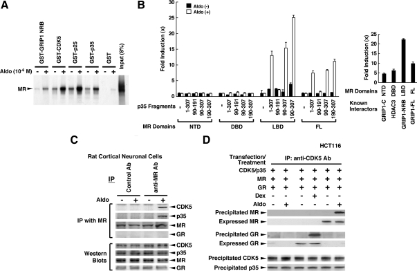
Glucocorticoids, major end effectors of the stress response, play an essential role in the homeostasis of the central nervous system (CNS) and contribute to memory consolidation and emotional control through their intracellular receptors, the glucocorticoid and mineralocorticoid receptors. Cyclin-dependent kinase 5 (CDK5), on the other hand, plays important roles in the morphogenesis and functions of the central nervous system, and its aberrant activation has been associated with development of neurodegenerative disorders. We previously reported that CDK5 phosphorylated the glucocorticoid receptor and modulated its transcriptional activity. Here we found that CDK5 also regulated mineralocorticoid receptor-induced transcriptional activity by phosphorylating multiple serine and threonine residues located in its N-terminal domain through physical interaction. Aldosterone and dexamethasone, respectively, increased and suppressed mRNA/protein expression of brain-derived neurotrophic factor (BDNF) in rat cortical neuronal cells, whereas the endogenous glucocorticoid corticosterone showed a biphasic effect. CDK5 enhanced the effect of aldosterone and dexamethasone on BDNF expression. Because this neurotrophic factor plays critical roles in neuronal viability, synaptic plasticity, consolidation of memory, and emotional changes, we suggest that aberrant activation of CDK5 might influence these functions through corticosteroid receptors/BDNF.


Figures

Figure 1
Figure 1
CDK5 suppresses MR-induced transcriptional activity. A, CDK5 suppresses MR-induced transcriptional activity in a kinase activity-dependent fashion. HCT116 cells were transfected with plasmids expressing the molecules indicated together with MMTV-Luc and pGL4.73-RLuc. Bars in the top panel represent mean ± se values of firefly luciferase activity normalized for Renilla luciferase activity in the presence and absence of 10−8m aldosterone (Aldo). Expression levels of MR and β-actin (bottom panel, top and bottom gel, respectively) in the Aldo-treated samples used in the top panel were examined with Western blots (bottom panel). *, P < 0.01; n.s., not significant between the two conditions indicated. B, CDK5 does not influence Aldo-induced nuclear accumulation of MR. HCT116 cells were transfected with indicated protein-expressing plasmids and were treated with 10−8m Aldo for 1 h. Total and nuclear MR were examined, respectively, in whole homogenates and nuclear extracts of these cells in Western blots by using anti-MR antibody. RLU, relative light units; WT, wild type.
Figure 2
Figure 2
CDK5 and MR interact with each other in vitro and in vivo. A, MR interacts with GST-fused CDK5, p35, and p25 in a ligand-dependent fashion in a GST pull-down assay. In vitro translated and 35S-labeled MR was incubated with bacterially produced and purified GST-fused proteins on GST beads in the presence and absence of 10−6m aldosterone (Aldo). Radiolabeled MR associated with GST-fusion proteins was visualized in SDS-PAGE gels; 8% of MR used for the reaction was loaded as input. B, MR interacts with p35 through its LBD in an Aldo-dependent fashion in a yeast two-hybrid assay. EGY48 yeast cells were transformed with p8OP-LacZ, pB42AD-p35 (1-307) -p35 (90-307), -p35 (1-191) -p35 (190-307), GRIP1-C, GRIP1-NRB, GRIP1-FL, or HDAC3, and pLexA-MR (1-984), -MR (1-602), -MR (602-670), or -MR (668-984) and incubated with 10−6m Aldo. The results of the interaction between MR domains and p35 fragments were shown in the left panel. The interaction of these MR domains in the presence of 10−8m Aldo with molecules already known to bind them is shown in the right panel to demonstrate the integrity of the expressed MR-related proteins. Basal β-galactosidase activities of MR NTD, DBD, LBD, and FL in the absence of their interactors are 33,009 ± 2147, 212 ± 9, 44,349 ± 983, and 69,662 ± 2862 (mean ± se, unit/OD600nm), respectively. Bars represent the mean ± se values of fold activation compared with baseline (left panel, the value obtained in the presence of pLexA, pB42AD-p35, and p8OP-LacZ and in the absence of aldosterone; right panel, the value obtained in the absence of interactors). C, Carboxyl terminal. Panel C, MR is associated with CDK5 and p35 in a ligand-dependent fashion in rat primary cortical neuronal cells. Rat primary cortical neuronal cells were incubated in the presence and absence of 10−8m Aldo for 1 h, and coimmunoprecipitation was carried out by using the antibodies (Ab) indicated. The results of coimmunoprecipitation are shown in the top four gels, whereas expression levels of CDK5, p35, MR, and GR are shown in Western blots in the bottom four gels by loading 10% of cell lysates used for the coimmunoprecipitation reactions. D, MR and GR, respectively, form a protein complex with CDK5 and p35 in a ligand-dependent fashion in HCT116 cells. HCT116 cells were transfected with the indicated protein-expressing plasmids and were treated with 10−8m Aldo or 10−6m dexamethasone (Dex) for 1 h. Coimmunoprecipitation was performed by using anti-CDK5 antibody, and precipitated MR, GR, CDK5, and p35 were examined (third top and bottom two gels). Expressed MR and GR were also shown in Western blots in second and fourth top gels by loading 10% of cell lysates used for the coimmunoprecipitation reactions. IP, Immunoprecipitation.
Figure 3
Figure 3
CDK5 regulates MR-induced transcriptional activity by phosphorylating the latter’s two serine (128 and 250) and one threonine (159) residues. A, A representative analysis of the phospho-MS of GST-hMR (1-602) phosphorylated by CDK5/p35. The peptide sequence analyzed in this panel is shown in the upper left corner. The serine phosphorylated by CDK5 is indicated with asterisk and underline. B, Serines 128 and 250 and threonine 159 are all necessary for CDK5 to suppress MR-induced transcriptional activity in HCT116 cells. Top panel, HCT116 cells were transfected with plasmids expressing the indicated MR mutants and CDK5/p35 together with MMTV-Luc and pGL4.73-RLuc. Bars represent mean ± se values of firefly luciferase activity normalized by Renilla luciferase activity in the presence and absence of 10−8m aldosterone (Aldo). *, P < 0.01, n.s., not significant between the two conditions indicated. Bottom panel, HCT116 cells were transfected with plasmids expressing the indicated MR mutants, and levels of MR proteins accumulated in the nucleus after treatment with 10−8m Aldo (top gel) and the control Oct1 (bottom gel) were examined in Western blots using their specific antibodies in the nuclear extracts obtained from these cells. C, CDK5 phosphorylates serines 128 and 250 and threonine 159 in an Aldo-dependent fashion in HCT116 cells. HCT116 cells were transfected with the indicated MR mutant-expressing plasmids together with CDK5- and p25-expressing plasmids and were treated with 10−8m Aldo for 1 h. MRs were immunoprecipitated by anti-MR antibody, and phosphorylated serine (top panel) or threonine (bottom panel) residues of MR by CDK5 were examined in Western blots by using antibody specific to phosphoserine or -threonine residues. D, CDK5 phosphorylates serine and threonine residues in an Aldo-dependent fashion in rat primary cortical neuronal cells. Rat primary cortical neuronal cells were treated with 10−8m Aldo and/or 20 μm roscovitine for 1 h. MRs were immunoprecipitated with anti-MR antibody, and serine- and threonine-phosphorylated MRs (top and bottom gel, respectively) were examined in Western blots by using antibody specific for phosphoserine or -threonine residues. WT, Wild type.
Figure 3
Figure 3
CDK5 regulates MR-induced transcriptional activity by phosphorylating the latter’s two serine (128 and 250) and one threonine (159) residues. A, A representative analysis of the phospho-MS of GST-hMR (1-602) phosphorylated by CDK5/p35. The peptide sequence analyzed in this panel is shown in the upper left corner. The serine phosphorylated by CDK5 is indicated with asterisk and underline. B, Serines 128 and 250 and threonine 159 are all necessary for CDK5 to suppress MR-induced transcriptional activity in HCT116 cells. Top panel, HCT116 cells were transfected with plasmids expressing the indicated MR mutants and CDK5/p35 together with MMTV-Luc and pGL4.73-RLuc. Bars represent mean ± se values of firefly luciferase activity normalized by Renilla luciferase activity in the presence and absence of 10−8m aldosterone (Aldo). *, P < 0.01, n.s., not significant between the two conditions indicated. Bottom panel, HCT116 cells were transfected with plasmids expressing the indicated MR mutants, and levels of MR proteins accumulated in the nucleus after treatment with 10−8m Aldo (top gel) and the control Oct1 (bottom gel) were examined in Western blots using their specific antibodies in the nuclear extracts obtained from these cells. C, CDK5 phosphorylates serines 128 and 250 and threonine 159 in an Aldo-dependent fashion in HCT116 cells. HCT116 cells were transfected with the indicated MR mutant-expressing plasmids together with CDK5- and p25-expressing plasmids and were treated with 10−8m Aldo for 1 h. MRs were immunoprecipitated by anti-MR antibody, and phosphorylated serine (top panel) or threonine (bottom panel) residues of MR by CDK5 were examined in Western blots by using antibody specific to phosphoserine or -threonine residues. D, CDK5 phosphorylates serine and threonine residues in an Aldo-dependent fashion in rat primary cortical neuronal cells. Rat primary cortical neuronal cells were treated with 10−8m Aldo and/or 20 μm roscovitine for 1 h. MRs were immunoprecipitated with anti-MR antibody, and serine- and threonine-phosphorylated MRs (top and bottom gel, respectively) were examined in Western blots by using antibody specific for phosphoserine or -threonine residues. WT, Wild type.
Figure 4
Figure 4
Aldosterone (Aldo) stimulates, whereas dexamethasone (Dex) suppresses, BDNF production of rat primary cortical neuronal cells. A, Aldo stimulates BDNF mRNA expression in rat primary cortical neuronal cells, whereas Dex suppresses it. Rat primary cortical neuronal cells were incubated in the presence of 10−8m Aldo or 10−6m Dex for 0, 1, 4, 10, or 24 h, and mRNA expressions of BDNF, TrkB, p75NTR, CDK5, and p35 were determined with SYBR Green real-time PCR. Circles and bars represent mean ± se values of mRNA expression of the molecules indicated corrected for that of RPLP0 in the presence of aldosterone (open circles) or dexamethasone (solid circles). B, Specific receptor inhibitors eplerenone and RU 486, respectively, abolish Aldo- and Dex-induced changes in BDNF mRNA expression in rat primary cortical neuronal cells. Rat primary cortical neuronal cells were incubated with the steroids indicated, and BDNF mRNA expression was measured with SYBR Green real-time PCR. Bars represent mean ± se values of mRNA expression of BDNF corrected for that of RPLP0. *, P < 0.01, compared between the two conditions indicated. C, Aldo increases, whereas Dex reduces, BDNF secretion into the culture medium of rat primary cortical neuronal cells. Rat primary cortical neuronal cells were incubated in the presence of 10−8m Aldo, 10−6m Dex, 10−6m eplerenone, and/or 10−5m RU 486 for the indicated time periods, and the concentrations of BDNF in medium were determined with a specific ELISA. Circles and bars represent mean ± se values of BDNF concentrations in the presence of Aldo (open circles) or dexamethasone (solid circles). *, P < 0.01, compared with the values in the presence and absence of eplerenone or RU 486 in the presence of same concentration of Aldo or Dex. D, Aldo, corticosterone (Cortico), and Dex differentially regulate BDNF secretion in rat primary cortical neuronal cells. Rat primary cortical neuronal cells were incubated in the presence of indicated concentrations of Aldo, Cortico, or Dex for 24 h, and the concentrations of BDNF in medium were determined with a specific ELISA. Circles/squares and bars represent mean ± se values of BDNF concentrations in the presence of Aldo (open circles), Cortico (open squares), or Dex (solid circles). *, P < 0.05; **, P < 0.01, compared with the values in the absence of steroids. E, Eplerenone and RU 486 differentially regulate Cortico-induced BDNF secretion in rat primary cortical neuronal cells. Rat primary cortical neuronal cells were incubated with the indicated concentrations of Cortico in the presence or absence of 10−6m eplerenone or 10−5m RU 486 for 24 h. Concentrations of BDNF in medium were determined with a specific ELISA. Squares/circles and bars represent mean ± se values of BDNF concentrations in the absence (open squares) or presence of eplerenone (solid circles) or RU 486 (solid circles). *, P < 0.05, compared with the values in the absence of eplerenone or RU 486.
Figure 5
Figure 5
CDK5 inhibitor roscovitine (Rosco) suppresses the stimulatory effect of aldosterone (Aldo) and the inhibitory effect of dexamethasone (Dex) on the BDNF production. A, Rosco suppresses the stimulatory effect of Aldo and the inhibitory effect of Dex on the mRNA expression of BDNF, whereas it weakly suppressed mRNA expression of p75NTR, but not TrkB, in rat primary cortical neuronal cells. Rat primary cortical neuronal cells were incubated with 10−8m Aldo or 10−6m Dex in the presence or absence of 20 μm Rosco for 9 h. mRNA expressions of the molecules indicated were determined by SYBR Green real-time PCR. Bars represent mean ± se values of mRNA expressions of the molecules indicated normalized for that of RPLP0 in the presence and absence of Rosco. *, P < 0.01; n.s., not significant compared with the values indicated or to the value obtained in the absence of steroids and roscovitine. B, Rosco suppresses the stimulatory effect of Aldo and the inhibitory effect of Dex on the BDNF secretion of rat primary cortical neuronal cells. Rat primary cortical neuronal cells were incubated with 10−8m Aldo or 10−6m Dex in the presence or absence of 20 μm Rosco for 24 h. Concentrations of BDNF in the culture media of these cells were determined with a specific ELISA. Bars represent mean ± se values of BDNF concentrations in the presence and absence of Rosco. *, P < 0.01; n.s., not significant compared with the values indicated or to the value obtained in the absence of steroids and Rosco. C, Knockdown of CDK5, but not of CREB1, suppresses the stimulatory effect of Aldo and the inhibitory effect of Dex on the BDNF secretion of rat primary cortical neuronal cells. Rat primary cortical neuronal cells were transfected with control, CDK5, or CREB1 siRNA and were incubated with 10−8m Aldo or 10−6m Dex for 24 h. Concentrations of BDNF in the culture media of these cells were determined using a specific ELISA. Bars represent mean ± se values of BDNF concentrations in the presence of indicated siRNA. *, P < 0.01; n.s., not significant compared with the value obtained in the presence of control siRNA and in the absence of steroids.

Similar articles

Cited by

References

    1. Kino T, Chrousos GP 2005 Glucocorticoid effect on gene expression. In: Steckler T, Kalin NH, Reul JMHM, eds. Handbook on stress and the brain. Amsterdam: Elsevier BV; 295–312
    1. de Kloet ER, Derijk RH, Meijer OC 2007 Therapy insight: is there an imbalanced response of mineralocorticoid and glucocorticoid receptors in depression? Nat Clin Pract Endocrinol Metab 3:168–179 - PubMed
    1. Fietta P, Fietta P 2007 Glucocorticoids and brain functions. Riv Biol 100:403–418 - PubMed
    1. Rashid S, Lewis GF 2005 The mechanisms of differential glucocorticoid and mineralocorticoid action in the brain and peripheral tissues. Clin Biochem 38:401–409 - PubMed
    1. Kino T, Chrousos GP 2004 Glucocorticoid and mineralocorticoid receptors and associated diseases. Essays Biochem 40:137–155 - PubMed

Publication types

MeSH terms